Surgical Management of Myopic Maculopathy: To Peel or Not to Peel the ILM?
Based on a presentation by Dr. Andrew Chang, Vitreoretinal Ophthalmologist and Surgeon, Medical Director, Sydney Retina Clinic, Consultant Vitreoretinal Surgeon and Head of Unit Sydney Eye Hospital, and Clinical Associate Professor, University of Sydney, Australia
Surgical management of myopic maculopathy remains one of the most unpredictable situations faced by vitreoretinal surgeons. In a lecture delivered at the recently held AVRTT@6 workshop in Ahmedabad, India, Dr. Andrew Chang led discussions and provided key insights into current approaches toward addressing the complications of surgery for myopic maculopathy.
According to Dr. Chang, the unpredictability of surgical management of myopic maculopathy arises due to several underlying mechanisms. Notably, there is marked inward vitreous traction and retinal arterial stiffening, resulting in failure of retinal stretching and non-compliance of the eye lens. Recent studies have discussed the classification of macular structure and the timing of surgical intervention. Further, Dr. Chang noted that in the early stages of disease characterized by foveoschisis, patients may be asymptomatic and currently there is no consensus on whether early surgical intervention might prove beneficial. In addition, he highlighted several unanswered questions concerning the surgical approach. These questions surround the safety of ILM peeling, outcomes of foveal sparing techniques and the effectiveness of triamcinolone. Furthermore, more clinical data is needed on the outcomes of the drainage of sub-retinal fluid via peripheral retinotomy or through the hole itself in cases of retinal detachment.
Dr. Chang discussed the surgical management of 2 cases of myopic maculopathy. The first case was a 35-year-old female with previous retinal detachment repaired with scleral buckling who presented with progressive worsening of vision and foveal detachment. Vitrectomy was performed and visual landmarks highlighted with triamcinolone. No internal limiting membrane (ILM) peeling was performed to avoid the risk of forming a macular hole. At postoperative day 10, patient was reviewed and the foveal detachment had settled, but patient later presented with an enlarging scotoma. A diagnosis of retinal detachment was made and the large volume of fluid was drained via a peripheral retinotomy.
The second case presented by Dr. Chang was that of a 43-year-old woman who presented with poor vision, a posterior staphyloma and foveal detachment. At surgery the fovea was difficult to visualize at the base of the staphyloma and triamcinolone was used (specifically Kenacort). At postoperative day 14, the patient presented with an enlarging scotoma and a diagnosis of macular hole retinal detachment was made. Drainage was performed though the macular hole. A few years later the patient presented again with schisis in the other eye associated with vitreal traction and was managed conservatively. Patient later presented with sudden loss of vision and a macular hole was identified. Intraoperatively, triamcinolone was used to highlight the visual landmarks, and standard ILM peeling and air-fluid exchange were performed. Schisis was resolved fully at postoperative review.
During the discussion session that followed, it was concluded that ILM peeling is useful in removing vitreal traction. Dr. Chang concluded that ILM peeling itself might increase the risk of macular hole retinal detachment. “As a safer option, foveal sparing ILM peeling, which avoids macular hole formation should be considered.”
The Versatile ILM and Macular Pathology
Based on a presentation by Dr. Shobhit Chawla, Medical Director and Chief Vitreoretinal Consultant, Prakash Netra Kendr, Gomtinagar, Lucknow, India
Surgical removal of the ILM represents one of the most important advances in vitrectomy in the past 15 years. In a lecture delivered at the recently held AVRTT@6 workshop in Ahmedabad, India, Dr. Shobhit Chawla shed light on recent advances in the understanding of the ILM structure, functionality and its role in macular pathology.
The ILM is a 1.2mm thick Periodic acid Schiff-positive structure. It is the boundary that establishes the contact and communication point of two compartments: the retina and vitreous. The sheer strength generated by the movement of the liquified vitreous induces specific inflammatory reactions on the ILM, while expression of contractile proteins leads to cellular transformation into fibroblasts, which induces scar tissue formation. Multiple pathologies can exist proximal and distal to the ILM, such as the posterior hyaloid phase, hyaline traction, partial thickness macular hole, and therefore, peeling the ILM resolves these multiple pathologies. ILM removal is performed to release the pathological influence of the vitreous on the retina, and is useful in restoring the normal anatomical shape of the macula and improving visual acuity. This technique has been utilized in a variety of indications such as diabetic maculopathy, retinal vein occlusion and retinal dysplasia.
However, ILM peeling can cause morphological changes at the macula like dissociated optic nerve fiber (DONF). Although no functional consequences have been attributed to this anatomical change, some studies have demonstrated that electrophysiological changes can occur after ILM peeling, and microperimetry may be useful in delineating functional outcomes of DONF. During his presentation, Dr. Chawla noted that a fundamental question in the field, is whether to peel the ILM or not. Currently, there are no answers to this important question. Though many surgeons opt to peel the ILM, peeling may not be necessary in absence of significant ILM damage.
Furthermore, caution is needed in the presence of circulatory and metabolic comorbidities.
Current data shows that epiretinal membrane (ERM) recurrence following combined ERM and ILM removal occurs in <9% of cases, and varies between 7.5-56%. Visual results have also been variable, and there is currently no consensus whether ILM alone, or the ILM and ERM, should be removed. Dr. Chawla explained that, given the variability of ERMs in clinical experience, consideration of ILM removal should be based on ILM appearance post-ERM removal at the time of surgery. Large areas of ILM are removed during ERM peeling, as evidenced by sequential staining and small punctate hemorrhages. Then Dr. Chawla provided more insights into surgical techniques of ILM peeling. He stressed the importance of removing all residual particles, to reduce incidence of postoperative fibrosis. In addition, Dr. Chawla advised the audience that the use of an inverted ILM flap technique for large, full thickness macular holes and myopic macular holes may be more effective than complete ILM peeling in cases of myopic macular holes with retinal detachment.
Myopic Foveoschisis: Looking Between the Layers
Based on a presentation by Dr. Hemanth Murthy, Consultant and Vice-president, Retina Institute of Karnataka, Bangalore, Karnataka, India
Myopic foveoschisis (MF), also known as myopic tractional foveoschisis, is increasingly being recognized as one of the major causes of visual loss in highly myopic eyes. Dr. Hemanth Murthy shared his experience in the management of MF at the recently held AVRTT@6 workshop in Ahmedabad, India. MF presents as pathological myopia in 8-24% of cases. Mechanically, MF is associated with tangential traction from the pre-macular membrane, and inverse traction from the posterior staphyloma.
According to Dr. Murthy, the etiology of MF is not known but it may be related to the relative thickness of the inner compared to outer retinal layers over the area of the staphyloma, the non-distensible nature of the ILM, and incomplete posterior vitreous detachment (PVD). The natural course is variable and patients may remain asymptomatic for long periods. Early symptoms could include blurring of vision, and progressively develop to foveal detachment and macular hole. In the presence of a lamellar hole, progression to retinal detachment is rapid. In discussing the surgical management of MF, Dr. Murthy utilized clinical cases and highlighted that the surgical approach involves vitrectomy and staining of the vitreous using triamcinolone combined with or without ILM peeling. Dr. Murthy concluded that in treating MF, ILM peeling ensures that cortical remnants are removed, but is slightly traumatic to the inner retina. Nonetheless, the results are excellent for the majority of patients. “Foveal sparing ILM peeling could be utilized if the fovea appears thin on OCT,” he added.
Vitrectomy for Endophthalmitis: Time is of the Essence
Based on a presentation by Dr. Andrew Chang, Vitreoretinal Ophthalmologist and Surgeon, Medical Director, Sydney Retina Clinic, Consultant Vitreoretinal Surgeon and Head of Unit Sydney Eye Hospital, and Clinical Associate Professor, University of Sydney, Australia
Endophthalmitis is still a rare but potentially eye-threatening complication of intraocular surgery. In a lecture delivered at the recently held AVRTT@6 workshop in Ahmedabad, India, Dr. Andrew Chang provided an overview of the current trends in the management of postoperative endophthalmitis.
While summarizing the landmark Endophthalmitis Vitrectomy Study (EVS) published in 1995, Dr. Chang stated the key historical importance of the EVS recommendations was the management of acute postoperative endophthalmitis. Notably, the study was the first to confirm the visual benefit of vitrectomy in patients presenting with acute postoperative endophthalmitis and visual acuity of light perception only. This was due to significantly improved outcomes of a 3X higher chance of achieving 20/40 vision or better and a reduced possibility of severe vision loss. However, if vision loss is at hand movement or better, then a tap or injection was the preferred approach. The study also showed that there was better prognosis in patients with better visual acuity scores and culture negative at presentation.
However, Dr. Chang questioned the relevance of said study findings in current practice, given the availability of better vitrectomy technologies, newer antibiotics and different procedures which were unavailable in the mid-1990s when the EVS was conducted. According to Dr. Chang, recent years have witnessed an exponential rise in the number of intravitreal injections (IVIs) in the management of cataracts and subsequently, an increased risk of endophthalmitis. However, studies have shown that the spectrum of organisms causing post IVI endophthalmitis differs from those implicated in following cataract surgery. Therefore, it is important to delineate the differences in post-IVI and post-cataract surgery endophthalmitis.
Dr. Chang and colleagues studied a consecutive series of 101 patients presenting with acute endophthalmitis. Out of those patients, 53 were post-IVI and 48 were post-cataract surgery. They found that post-IVI endophthalmitis was associated with poorer visual outcomes, and this was related with an increased chance of streptococcal infection. Furthermore, streptococcal infections were associated with increased odds of enucleation.
There are several advantages of early vitrectomy, Dr. Chang noted, such as better diffusion of antibiotics, earlier visualization of the retina and debridement of bacteria, inflammatory debris and toxins. However, there are challenges including iatrogenic retina damage and anesthetic risks. To assess the efficacy and safety of early vitrectomy in acute-onset endophthalmitis, Dr. Chang and colleagues at the Sydney Eye hospital conducted the Early Vitrectomy Study. This was a retrospective study of 64 consecutive patients from 2009-2013, who developed endophthalmitis following a range of procedures. Patients were followed and the primary outcome was visual acuity (VA) at 12 months. In these patients, immediate vitreous tap and injection of antibiotics were performed under local anesthesia in the emergency units. Following this, patients underwent 23-G vitrectomy within 72 hours, with anterior chamber washout, corneal debridement, core vitrectomy and intravitreal vancomycin. The mean age of patients was 77.5 years and mean time from onset of inciting procedure was 5.7 days. In majority of patients, the inciting procedure was phacoemulsification and IVI, while the most predominant bacteria species implicated were streptococci and S. epidermidis.
Overall, the study findings were impressive. Visual outcomes were improved in 89% of patients and 42% had final VA better than logMAR 0.477 (Snellen 6/18). VA was better in post-cataract surgery as compared to post-IVI. Retinal detachment occurred intraoperatively in 9.4% of eyes, and postoperatively in 6.2%, and epiretinal membrane formation occurred in 9.4%. Based on their findings, Dr. Chang concluded that micro-incisional vitrectomy is associated with better visual outcomes and effective in improving vision in acute infective endophthalmitis (culture negative) and post-cataract surgery endophthalmitis. In addition, visual prognosis is limited to macular pathology.
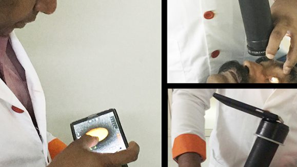
Editor’s Note: AVRTT@6, the 6th Advanced Vitreo-Retinal Techniques & Technology event took place on March 17 -18, 2018, at Narayani Heights, Ahmedabad, India. AVRTT@6, is a live surgery workshop organized by the Retina Foundation & Eye Research Center, headed by Dr. Manish Nagpal. Reporting for this story also took place at AVRTT@6.