Prior to its debut into ophthalmic imaging in the late 1990s, adaptive optics (AO) was a highly-utilized method of reducing atmospheric distortion in astronomy. Astrophysicists rely on AO to help reduce granular image artifacts, minimize blur and sharpen faint features of objects from the cosmos through their telescopes.
We are similarly familiar with the granular artifacts that distort or obscure microscopic details of optical coherence tomography (OCT), which despite decades of technological improvements, remain a challenge of the imaging modality. As OCT technology has advanced beyond time-domain, spectral-domain and most recently swept-source OCT, the capabilities to visualize high resolution cross sections of the retina have revolutionized ophthalmic care.
OCT is, however, restricted to the axial direction and the monochromatic aberrations limit transverse or lateral resolution, reducing overall image quality. By pairing AO technology with OCT, microscopic retinal structures that were previously unable to be seen can now be examined. AO technology can adjust for, and limit the eye’s optical aberrations, which are responsible for much of the blurring and image artifacts. With AO, the optic nerve head (ONH) and retina can be imaged in three dimensions, visualizing discrete structures, such as photoreceptors, retinal nerve fiber layer (RNFL) bundles, and the lamina cribrosa.
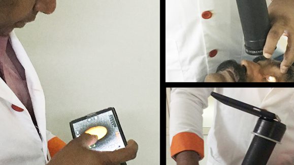
In a recent issue of Progress in Retinal and Eye Research, Zachary Dong and colleagues at the University of Pittsburgh Medical Center Eye Center and New York University Langone Eye Center reviewed the history and application of AO to ophthalmic imaging, specifically to glaucoma diagnosis and management. They discuss AO-OCT and its use in combination with other modalities, such as scanning laser ophthalmoscopy (SLO) and AO fundus cameras. One of the study authors, Dr. Gadi Wollstein commented that, “adaptive optics is a promising technology for enhancing image quality that has important impact of identifying small features in the eye and advance understanding of disease processes and progression.”
Imagine having a clear view into the retinal microstructures, quantifying blood flow and capillary density, and measuring RNFL bundles – all of which are currently possible with AO. This level of detail is opening an entirely new world to clinicians and researchers…but what exactly can we see, and what does it all mean?
Although AO-OCT provides the ability to look deeper and more clearly into smaller structures, the instrumentation does have the disadvantage of having a smaller field of view, restricting imaging to specific regions and identifying such regions can be challenging. Also, determining which of these observations are characteristic of different disease states remains complicated. The existence of reference databases that clearly identify findings of interest, comparing healthy to clinically relevant characteristics, are still lacking.
There is no doubt that OCT has improved glaucoma diagnoses and management. With the addition of AO image analysis to the research arsenal, assessment of the functionality of the retinal microstructure will lead to further understanding of disease pathogenesis and outcomes. AO-OCT has demonstrated the ability to evaluate the structural characteristics of the RNFL and identify potential risk factors implicated in glaucomatous ONH damage. The expectation is that new pathogenic events will be uncovered, leading to earlier diagnosis and new treatment targets.
When asked about the future of AO and its role in the clinical setting, Dr. Wollstein stated, “the technology right now has limited clinical use because it is cumbersome and expensive but with the advancement of technologies it is very likely to have an important role in clinical management in the future.”
Reference:
Dong ZM, Wollstein G, Wang B, et al. Adaptive optics optical coherence tomography in glaucoma. Prog Retin Eye Res. 2016;S1350-9462(16)30064-7.