Since its first commercial introduction in 1996, optical coherence tomography (OCT) has been described as the most valuable advancement in retinal diagnostic imaging in nearly half a century. The prior ‘ground-breaking’ advancement had been fluorescein angiography in the late 1950s.
Now, we find ourselves entering a new and exciting era of retinal imaging with the introduction of OCT angiography (OCTA) technology, providing the ability to non-invasively visualize retinal blood flow in a matter of seconds without any need for a contrast medium.
With so much innovation happening in OCTA, there is an increasing interest not just in North America and Europe, but in Asia-Pacific as well.
A crash course in OCTA
Unlike the static images obtained with traditional OCT, OCTA uses a series of sequential scans, from a single cross-section of the retina. It is important to note that the scans have to be from the same spot of the retina, otherwise the OCTA algorithms will not provide the desired flow information. The scans are compared against one another and the differences indicate the presence of motion or blood flow. It uses a principle of motion contrast, resolving the signal of the blood flow, a movement within a motionless structure. Within seconds, clinicians can clearly observe areas of perfusion, or non-perfusion. Although OCTA does not deliver exactly the same level of detail as the traditional 2D perspective of fluorescein angiography, the clinical information provided is unsurpassed. The 3D images that are produced by OCTA can be separated into individual layers of retinal vasculature, delivering an unprecedented level of detail and the ability to isolate areas of interest. Imaging of the retinal and choroidal vasculature reveals new details about blood flow, measurements of choroidal neovascularization, and further visualization of the peripapillary network. Additionally, there may be an opportunity for application of OCTA in glaucoma diagnosis and management, as perfusion of the optic nerve has been associated with the disease.
Three leading OCTA systems involved in this breakthrough technology include the AngioVue® System from Optovue, CIRRUS™ HD-OCT AngioPlex™ from ZEISS Medical Technology, and the OCT Angiography Module for Heidelberg Engineering’s popular SPECTRALIS® system. While this list may not be exhaustive, it’s a good start into understanding commercial offerings in this exciting new OCT world.
Optovue’s AngioVue® Imaging System
Optovue, based in Fremont, California, was the pioneer and very first provider of OCTA technology, introducing the world’s very first commercially available platform in 2014. The AngioVue Imaging System uses SSADA (Split-Spectrum Amplitude Decorrelation Angiography), developed by David Huang, M.D., Ph.D., at Oregon Health and Science University. SSADA is a proprietary algorithm that shortens the scan time needed to acquire the sequential B-scans needed for OCTA acquisition, while maintaining the highest image quality by optimizing the signal to noise ratio (SNR). Basically, it’s elevated image quality in a shorter amount of time. There are already well over 200 peer-reviewed publications detailing the AngioVue Imaging System in clinical applications. The AngioVue System is a comprehensive ophthalmic platform, also addressing clinical imaging needs, including the front of the eye and the optic nerve.
In June of 2016, Optovue announced the availability of DualTrac™ Motion Correction technology for use with both the AngioVue and AngioVue Retina systems. DualTrac is a two-level approach to correcting motion artifact on patients who have difficulty focusing on a central visual point during an eye exam, essential for high-quality OCTA imaging. The first level, real-time tracking, provides a correction for any of the naturally occurring blinking or involuntary eye movements that can negatively impact image quality. This means that patients can still blink during image scanning, preventing the need for multiple repeat scans, reducing burden to patient and clinic staff. The second level occurs during post processing and provides pixel level correction for finer eye movements such as saccades.
“We use AngioVue OCTA for our patients who need angiography, as a first step before considering more invasive options such as fluorescein and indocyanine green angiography. Often, it gives us an answer without having to resort to dye-based angiography, and has reduced the amount of time and discomfort our patients are subjected to because it is completely non-invasive,” explained Dr. Nadia Waheed, Director of the Boston Image Reading Center and Associate Professor of Ophthalmology at the Tufts University School of Medicine in Boston.
“I have witnessed the number of fluorescein angiography procedures decrease because we now have a non-invasive option. Additionally, we are able to repeat it as frequently as we like, so it gives us a great way to monitor disease progression and response to therapy,” she added.
The AngioVue system is available in two different configurations, depending on the needs of the practice. AngioVue Retina is optimized for the retina practice, combining functional OCTA with structural OCT scans, and the more comprehensive AngioVue system includes OCTA capabilities and structural OCT for retina, optic nerve and anterior segment applications.
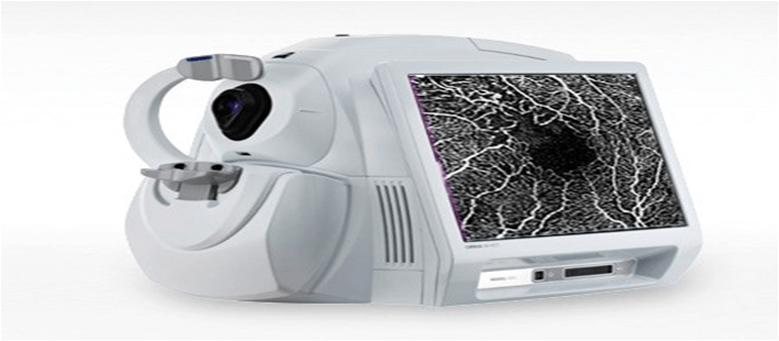
ZEISS AngioPlex™
AngioPlex™ from ZEISS was the very first OCTA system to receive U.S. Food and Drug Administration (FDA) clearance and is available on the CIRRUS™ HD-OCT 5000 platform, allowing ophthalmic practices the flexibility to easily integrate this novel vascular imaging application with their standard OCT diagnostic imaging. Different than the other OCTA systems that typically require multiple OCT scans to generate one single OCT angiography image, ZEISS AngioPlex allows for the visualization of both vascular and structural information from a single non-invasive scan. ZEISS AngioPlex uses a real-time retinal tracking system, FastTrac™, which actively eliminates eye motion, delivering motion-artifact-free images of the perfused retina. Additionally, FastTrac enables follow-up OCTA images to be taken at precisely the same location as at previous visits, in order to assess treatment efficacy and monitor disease progression.
Describing his experience with the AngioPlex technology, Dr. Deepak C. Bhatt, M.D., from the UBM Institute in Mumbai, said: “ZEISS AngioPlex is a comprehensive imaging tool for imaging of the macula, including OCT and OCT Angiography. The resolution of B Scan OCT and angiography are excellent. The acquisition time is short and the FastTrac system within AngioPlex are the highlights of the AngioPlex system; it works extremely well for patients with poor fixation.” In order to achieve the ultra-clear vascular images, ZEISS AngioPlex is powered by Optical Micro Angiography (OMAG) Algorithms, which is an image processing technique that takes full advantage of not only amplitude but also phase OCT signal data to deliver the highest-quality OCTA images.
The full depth color encoded images using AngioPlex clearly illustrate vascular pathology. “The color coding of all three retinal vasculature and projection removal software works well in practically all situations,” explained Dr. Bhatt. In diabetic retinopathy, it can show microaneurysms and areas of ischemia. In age-related macular degeneration it can reveal the presence of a choroidal neovascular membrane. And in vascular occlusions it can show the location of the occlusion and the extent of the ischemia.
Heidelberg Engineering’s OCTA Module for SPECTRALIS® Imaging Platform
Heidelberg Engineering has had an OCTA module for their popular SPECTRALIS® imaging platform available to customers outside the United States since late 2016. The expandable diagnostic platform allows for high resolution 3D, layer-by-layer examinations of the perfused retinal and choroidal vascular networks, using a complex mathematical model fed by full spectrum OCT data. The mathematical model is highly sensitive, allowing for visualization of the smallest of changes between consecutive OCT scans, representing the vascular flow and resulting in high contrast between areas of vascular flow and surrounding tissue.
Because of the multimodal imaging capabilities of SPECTRALIS, clinicians can integrate and compare OCTA to other modalities such as structural OCT and dye- based fluorescein (FA) or indocyanine green (ICGA) scanning laser angiography for a more comprehensive examination. This hybrid angiography approach provides the opportunity for OCTA images to be directly correlated, pixel to pixel, to FA or ICGA, setting a baseline for follow-up, OCTA-only examinations.
As with other SPECTRALIS modalities, the OCT Angiography Module also uses the TruTrack Active Eye Tracking technology that limits motion artifacts and ensures high resolution images. This OCTA delivers a lateral resolution of 5.7 microns per pixel for visualization of capillaries, and an axial resolution of 3.9 microns per pixel for precise multilayer segmentation.
As the world’s population ages and retinal vascular diseases remain leading causes of global blindness, OCTA will undoubtedly have a significant clinical impact. In many cases, and for specific data, dye-based FA will still have to be performed, but it is reasonable to believe that because of its non-invasive nature OCTA will be used at times when FA/ICGA would have not been indicated, providing additional diagnostic information and reducing the burden for patients and ophthalmic staff. There is no doubt that OCTA delivers an exciting advancement in clinical and imaging diagnostics, although research is ongoing related to the standardization of image generation, quantification and interpretation of findings.
“OCT angiography is a new imaging tool and we are still in the learning curve,” explained Professor Giovanni Staurenghi, from the University of Milan.
Discussing its advantages and pitfalls is an important and honest way to approach this new angiography and we have shown that it is advisable to exercise caution when using new technology that we do not necessarily fully understand yet. We know that using multimodal imaging provides a better diagnosis than using single imaging modes alone and it seems OCT angiography will play an important role in multimodal imaging in the future,” he added.