Retinal vein occlusion (RVO) is one of the most common causes of retinal vascular disease, with an estimated worldwide prevalence of 16 million patients globally (0.5%). Occlusion of the retinal vein leads to a decrease in retinal circulation outflow which may result in ischemia, macular edema, retinal damage and ultimately blindness.
Although lasers have successfully been used since the 1970s to treat these sequelae of vein occlusions, anti-VEGF agents are increasingly being used as first-line treatment for vision loss for this disorder. Central and branch retinal vein occlusions have different pathophysiologic mechanisms which must be addressed separately. Vitreoretinal surgeon, Prof.
Peter Stalmans, MD, PhD, from the Department of Ophthalmology, UZ Leuven, Belgium, presented on the topic of “Robot-Assisted Retinal Vein Cannulation for Retinal Vein Occlusion” during the APVRS 2017 meeting held in Kuala Lumpur, Malaysia.
Retinal vein cannulation (RVC) is a difficult procedure performed for drug delivery into small retinal veins. The currently available glass cannulas used in this procedure are hard to visualize and fragile, making the procedure a challenging one, even for the most experienced of surgeons. Until now, robotics have never been used in RVC.
Application of Robotics in RVC
Prof. Stalmans and the Department of Mechanical Engineering at KU Leuven began to explore possible options for the application of robotics in RVC as early as 2010. Several robotic prototypes were developed and tested. A major achievement of this process was the development of an ultrathin aluminum coated glass microneedle with a 30-micron outer diameter for the delivery of a thrombolytic agent (ocriplasmin) into the retinal vein. Pre-clinical, in vivo results demonstrated the technical capabilities of performing a prolonged retinal vein cannulation using robot-assisted RVC with a 73.3% complete success rate, and no technical failures with the device when performed on 9 sets of pig’s eyes.* In 5 of the eyes, a second cannulation at the optic disc border was performed which resulted in an average cannulation time of 361.8 (± 138.5) seconds.
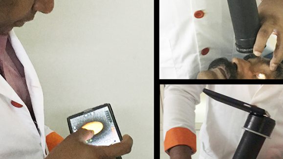
Dr. Stalmans performed the first ever robot-assisted RVC in a human patient with central retinal vein occlusion (CRVO) in January 2017. With the aid of a robotic co-pilot, up to 5 mg of ocriplasmin (Jetrea, ThromboGenics, NJ, USA) was infused over a maximum of 10 minutes into the occluded vein using a needle measuring 0.03 mm in diameter. This was part of the Phase 1 trials which had a total of 4 patients. Results of the phase 1 trials investigating the feasibility and safety of a robot-assisted RVC in these 4 patients with CRVO were recently published which demonstrated the technical success in the cannulation of the retinal vein and delivery of ocriplasmin with an average infusion time of 355 seconds per case.
CRVO can either take on a mild, nonischemic form or a severe, ischemic form with a much worse visual prognosis. The best predictor of final visual acuity is the vision at presentation with 80% of patients presenting with worse than 20/200 vision unable to achieve improvement. The etiology
of this condition is thought to be thrombosis of the central retinal vein posterior to the lamina cribrosa sclerae. Risk factors include uncontrolled diabetes, hypertension and open angle glaucoma. Occasionally, patients with clotting disorders, dysproteinemias and vasculitis can develop CRVOs. Following the inciting event, an average of 16% of all patients develop neovascularization of the iris and/or angle which can cause difficult to manage neovascular glaucoma.
MYNUTIA, a project of the Department of Mechanical Engineering at the KU Leuven created in close collaboration with the KU Leuven Technology Transfer Office, is aiming at commercializing this promising technology by stretching the limits of eye surgery with state-of-the-art robotic technology. With the aid of its surgical robots, eye surgeons can improve the quality of existing therapies and perform previously impossible treatments that have the potential to give millions of visually impaired and blind people a chance to regain their vision.
A Unique Stabilization System
In contrast to traditional surgical robots, no joystick is required to operate the robot. This surgical procedure designed by Leuven engineers and clinicians allows the surgeon to work in co-manipulation with the robot by holding the instrument as a normal vitreoretinal surgeon would during routine surgery. For maximum safety and intuitiveness, the surgeon retains direct control of the instrument and its motion, guiding it whilst the robot focuses exactly on what matters most: increasing the surgical precision over ten times by eliminating vibrations and stabilizing the movements. This precision enhancement is obtained by offering stabilization on three different levels: the surgeon’s hand, the patient’s eye and the surgical instrument.
Once the needle is successfully positioned in the retinal vein, the robot is locked and both the eye and the needle are automatically held in place. Delivery of desired drug into the vein is then achieved.
For the Phase 1b trials in 2018, improvements in visualization with a Rescan 700 microscope (Carl ZEISS Meditec, Jena, Germany) and enhancement of the robotic device which now has improved ergonomics and stability will be tested alongside intraoperative iOCT and angiography.
References:
APVRS 2017 Meeting Presentation by Prof. Peter Stalmans, Department of Ophthalmology, UZ Leuven, Belgium
Willekens K, Gijbels A, Schoevaerdts L, et al. Robot-assisted retinal vein cannulation in an in vivo porcine retinal vein occlusion model. Acta Ophthalmol. 2017;95(3):270-275.