Sponsored by Optomed
Accessibility, quality optics and AI combine to enable oculomics in Optomed’s newest handheld fundus camera Optomed Lumo
Not only is retinal imaging critical to detect eye disease, studies show it can also be used to predict other systemic conditions. Growing evidence indicates that the eyes can reveal more about overall health than just ocular disorders. This is because the eyes’ structure and function can mirror symptoms of other health conditions, including cardiovascular disease, neurodegenerative disorders and kidney problems.1
Positioning the eye as a window into the wider health landscape has given rise to oculomics, which uses ophthalmic biomarkers to understand mechanisms, and detect and predict systemic disease. Oculomics has been enabled by the widespread clinical adoption of high-resolution and non-invasive imaging, the availability of large studies and data sets, and the development of novel analytical methods like artificial intelligence (AI).1
READ MORE: Is This Canada-Approved Retinal Oximeter The Next Step for Clinical Oculomics?
This better understanding of the relationships between the body and the eye, along with developments in both diagnostics and AI, could help predict the risk of systemic disease and create more opportunity for prevention.1 According to Zhu et al.,”these innovations hold the potential to significantly enhance diagnostic accuracy, improve patient outcomes and offer novel insights into the interplay between ocular and systemic health.”1
Where oculomics meets innovation
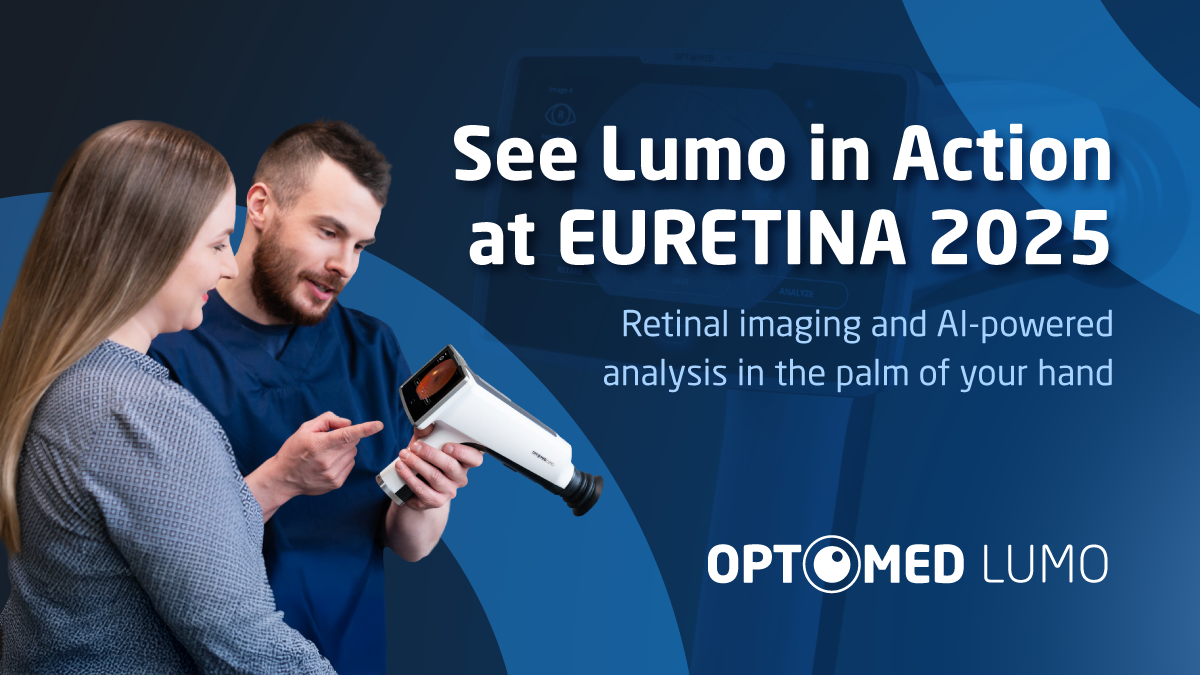
One recent advance in retinal imaging is particularly well-suited to provide a platform for disease detection and ultimately, oculomics: The Optomed Lumo®, an AI-powered, handheld fundus camera. The Finnish company’s previous handheld fundus cameras’ diagnostics are supported by clinical evidence2-4 and backed by 20 years of innovation experience and 40 international technology patents.
READ MORE: FDA Clears Optomed’s Handheld AI Screening Device for Dr in the US
Indeed, the Optomed Lumo was created on the shoulders of giants: Its predecessor Aurora AEYE is the only handheld fundus camera with FDA-clearance for autonomous diabetic retinopathy (DR) detection, with clinically validated image quality that’s equivalent to desktop camera images and suitable for screening. Now, it’s time for Optomed Lumo to take this image quality a step further, with a new high-sensitivity, low-noise sensor.
Let’s dive deeper into Optomed Lumo’s characteristics to see how they promote quicker detection, more reliable diagnostics and provide a platform for oculomics.
Screen patients anywhere
Reaching patients where they are opens up new avenues of care for those with reduced access to healthcare. The portability of Optomed Lumo enables patient screening beyond the ophthalmology clinic setting—whether in large-scale or mobile screening initiatives, or within emergency and pediatric departments.
It has a new intuitive user interface that enables any health professional to use the camera which provides a fast and effective tool for different consultation pathways. It also adapts to various healthcare settings and can be used as a handheld device or attached to a base for stationary use.
Increased accessibility coupled with the emerging field of oculomics means that more patients could receive sight- and life-saving diagnostic care.
WATCH NOW: A Chat About Optomed Aurora® IQ With Dr. Rudrani Banik
Better detection with high quality optics
Optomed Lumo makes early retinal changes more visible and easier to detect thanks to Optomed’s High-Contrast Optical Design™. Using optical filtering to enhance the contrast of retinal features, High-Contrast Optical Design™ results in high quality images that help support early and accurate diagnosis.
In addition, the Optomed Lumo is non-mydriatic, features a 50° field of view and produces images with 12 megapixel resolution. The camera is also user-friendly for operators, featuring a 5-inch touchscreen, programmable imaging sequences, Smart Autofocus™ and intuitive user guidance.
Collectively, the camera’s features create images that can be used to detect and diagnose retinal disease.
Connectivity and AI-powered analysis
These days connectivity is key, in settings both in- and outside of the clinic – and the Optomed Lumo’s AI integration means that it can easily adapt to various clinical needs and workflows.
The Optomed Lumo can complete data transfer and AI analysis on the spot, delivering results instantly to the camera and Optomed Portal. It can seamlessly integrate images with hospital systems using a direct DICOM connection. It also features USB and WLAN connections for image transfer, along with cloud and mobile device connectivity to make sharing results simple.
Beyond connectivity and diagnostic support, the Optomed Lumo can be integrated with different AI algorithms to detect disease, and AI-screening can be completed in one minute, making screening fast and effective.
READ MORE: Seeing the Unseen: Adaptive Optics Enters the Clinical Arena
Applying AI in the real world
Efficient screening and improved treatment of both diabetes and its visual counterpart DR have improved the visual prognosis for the disease.5 But as the number of diabetic patients continues to increase, the need for accessible and automated solutions to alleviate the burden on both patients and providers also grows.
In a study5 by Kubin et al., the authors shared that “implementation of telemedicine solutions, mobile handheld devices and AI-based automated analysis for DR might help alleviate the burden for screening and improve cost-effectiveness.”
To learn more about AI’s ability to detect DR, the Optomed Aurora was used to reveal how 21 algorithms performed in a real-world setting. In total, 312 eyes of 156 patients were graded by experienced ophthalmologists and the 21 AI algorithms. Of the 21 AI algorithms, 19 (90.5%) resulted in grading for DR at least in 98% of the images. The authors concluded that “fundus images captured with Optomed Aurora were suitable for DR screening.”
READ MORE: World-First Comprehensive Government-Backed AI-Powered Eye Screenings Go Live in Kerala
The most recent real-world study6 by Huhtinen et al. demonstrated the exceptional diagnostic performance of Aireen AI algorithm combined with Optomed Aurora fundus imaging for DR detection, achieving 94.8% sensitivity, 91.4% specificity and 92.7% diagnostic accuracy.
According to the authors, “The validated use of AI with a handheld fundus camera may streamline the screening process, reduce the burden on health care professionals, and improve access to screening and patient outcomes through enhanced diagnostic accuracy.”6
Using AI doesn’t only expand access to eye screening, it can also empower early detection and predictive healthcare interventions, transform eye screening into a powerful diagnostic tool, and to enable health detection from the eye. All of which aligns with oculomics and Optomed’s vision for improved patient outcomes.
Visit the Optomed booth (#3.A68) at EURETINA 2025 to learn more about the new Optomed Lumo AI-powered handheld fundus camera and how it facilitates the advancement of oculomics with more accessible and earlier disease detection.
Editor’s Note: The 25th EURETINA Congress is being held from 4-7 September, in Paris, France. This content is intended exclusively for healthcare professionals. It is not intended for the general public. Products or therapies discussed may not be registered or approved in all jurisdictions, including Singapore.
References
- Zhu Z, et al. Oculomics: Current concepts and evidence. Prog Retin Eye Res. 2025;106:101350.
- Midena E, et al. Handheld Fundus Camera for Diabetic Retinopathy Screening: A Comparison Study with Table-Top Fundus Camera in Real-Life Setting. J Clin Med. 2022;11(9):2352.
- Kubin AM, Wirkkala J, Keskitalo A, Ohtonen P, Hautala N. Handheld fundus camera performance, image quality and outcomes of diabetic retinopathy grading in a pilot screening study. Comparative Study Acta Ophthalmol. 2021;99(8):e1415-e1420.
- Midena E, Zennaro L, Lapo C, Torresin T, Midena G, Frizziero L. Comparison of 50° handheld fundus camera versus ultra-widefield table-top fundus camera for diabetic retinopathy detection and grading. Eye (Lond). 2023;37(14):2994-2999.
- Kubin AM, Huhtinen P, Ohtonen P, Keskitalo A, Wirkkala J, Hautala N. Comparison of 21 artificial intelligence algorithms in automated diabetic retinopathy screening using handheld fundus camera. Ann Med. 2024;56(1):2352018.
- Huhtinen P, Kubin AM, Dvořák K, Sliva M, Bayer J, Hautala N. Real-World Evaluation of Artificial Intelligence-Based Diabetic Retinopathy Screening Using the Optomed Aurora Handheld Fundus Camera. Diabetes Technol Ther. 2025 Aug 18. [Epub ahead of print.]
