We all have our weaknesses and this article is going to (rather unexpectedly, it must be said) start with a story about one. Your writer has a peanut allergy: It’s relatively minor considering he can be in the presence of them and not immediately run into problems, but still strong enough to make him jumpy with new food and obsessively check menus. On this occasion, he was meeting a friend for lunch — some vegan dim sum (which as a carnivore he was already suspicious of) — and ordered some tofu dumplings with spicy sauce.
He asked about the presence of nuts in English, his friend did the same in the local language, and was reassured there were none. Turns out, there were, and a pleasant afternoon turned into an impromptu trip to the hospital. A few hours later after having his body pumped full of steroids, antihistamines and a number of the drugs he couldn’t pronounce, he left feeling woozy, craving sugar with a wallet some $10 or so lighter (the benefits of being European).
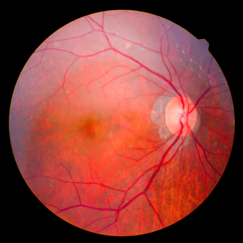
The point is, we all have our weaknesses and in our writer’s case, peanuts are certainly one of them, but perhaps what’s more important is how medicine has changed to treat said weaknesses over the years. A relatively mild reaction to peanuts in 2021 is far easier to deal with than it would have been 200 years ago — and as he also has asthma, your correspondent is especially glad he wasn’t alive 2,000 years ago. The leaps and bounds medical science has made in treatment over the centuries are remarkable, and indeed the last 100 years in particular, witnessed incredible progress in posterior segment treatment.
Looking Way Back with Hippocrates
We can point to the Greek physician Hippocrates as being the most likely candidate to indicate the posterior segment. Based on the findings of Alcmaeon of Croton, Hippocrates points to it comprising three layers: 1) a thick outer layer, 2) a thinner middle layer which may protrude like a bladder when injured and 3) an inner layer which is thinnest and very prone to damage. His work would be expanded on by fellow Greek Herophilus, whose surviving fragments show his understanding of the eye to include the lens (a drop-like body named crystalloids), and a large empty space (locus vacuus) containing humour, likely representing the anterior chamber.1
It wouldn’t be until the golden age of ophthalmology in the 19th century — and the invention of the ophthalmoscope by German physician Hermann von Helmholtz, which allowed doctors to fully examine the inner eye — that posterior segment treatment became fully differentiated.1
Now, we live in an era of constant technological advancements (not just in ophthalmology), and progress can come at breakneck speed. We thought: “Well, we know a pretty good deal about the posterior segment, but who knows better than an ophthalmologist?” So, we reached out to some of the posterior segment’s rockstar doctors to ask them about their history working in the field, and how much it has changed.
Drastic Improvements in Macular Hole Surgery

“I started as an ophthalmologist around 25 years back … and as you would expect, a lot has indeed changed in the field of the posterior segment. From diagnostics and imaging, to surgical decision-making and instrumentation, almost everything has changed for the better,” said Dr. Manish Nagpal, a vitreoretinal specialist and director at the Retina Foundation in Ahmedabad, India.
“When I started out, macular hole was considered to be an untreatable condition, however, today one can achieve an almost 95% success rate with surgery for the same condition. Vitreomacular traction is also diagnosed and understood better due to improved imaging, and therefore can be operated as indicated with more accuracy,” said Dr. Nagpal.
A macular hole is a small break in the macula and can cause distorted central vision and blurring. The condition is usually caused by shrinking vitreous due to aging, and as it pulls away from the retinal surface, vision begins to degrade. Natural fluid fills the area where the vitreous has contracted and some patients experience no issues during this process. However, if the vitreous is firmly attached to the retina when it pulls away, it can tear the retina and create a macular hole.2
Macular hole is a relatively common condition and for years it was thought to be untreatable because of missing tissue and photoreceptor degeneration. Initial attempts to treat the condition involved the use of laser photocoagulation; however, it would be the advent of keyhole surgery and improved imaging techniques that would allow this common, yet destructive, disease to be fully treated.3
Now, many retinal surgeons are able to treat macular hole via macular hole surgery, a form of keyhole surgery performed under a microscope. In this process, the vitreous jelly is removed and the inner limiting membrane peeled away, allowing a temporary gas bubble to be pressed into the hole flat to help it seal. The use of this technique resulted in remarkable improvement in patient outcomes, and nowadays, most patients who present less than six months from symptoms onset report a full recovery.
“This was a condition where the patient was certain to lose their central vision with time; today, we can save that vision,” Dr. Nagpal said.
And it’s not just keyhole macular surgery that is being used to cure this one-time vision killer. According to Dr. Hudson Nakamura, a retinal specialist at the Brazilian Center for Eye Surgery in Goiania, Brazil. He points to the use of the internal limiting membrane (ILM) technique as being a great discovery in macular hole treatment.
“ILM peeling for macular hole is fast on the way to becoming the tested and established procedure in the field. That’s just one example besides the comparative results involving the use of this technique from combined procedures such as scleral buckling surgery and vitrectomy,” Dr. Nakamura said.
“Also, with the development of minimally invasive vitreoretinal surgery, surface diseases became a lot better understood. This is because when smaller instrumentation took over for larger gauges, we could manage better and have greater outcomes with surgeries involving macular holes,” he added.
Leaping forward with Smaller Gauges
While speaking with Media MICE, Dr. Nakamura was keen to stress the importance of the improvement in gauges in vitreoretinal surgery, which allows for exponentially smaller incisions to be made in the ocular surface. He described that when he started out, the 20-Gauge was used for vitrectomies, and for some procedures a 19-Gauge was used to extract intraocular foreign bodies (IOFBs). The picture he describes today through his experience as a clinician is markedly different, with much more choice now available.
“In 2000 I started my practice with minimally invasive vitreoretinal surgery (MIVS) and it was great to see that from the development of the smaller gauge instruments,” Dr. Nakamura said.
“This began with the 23-Gauge, which was first developed for vitrectomy by Peyman back in 1990 for biopsy purposes; then the 25-Gauge variant for transconjunctival vitrectomy was used back in 2002, by Fuji et al. Subsequently, Eckart developed the 23-Gauge transconjunctival vitrectomy as an alternative to the 25-Gauge system, followed by the development of 27-Gauge by Oshima,” he said.
The Futuristic World of Gene Therapy and Implants

Retinitis pigmentosa (RP) is another condition that has exemplified the recent rapid advances in posterior segment treatment, said Dr. Nakamura. This condition is usually a single gene disorder, while diabetic retinopathy has a multifactorial (also known as polygenic or complex) etiology. The prevalence of RP is estimated 1 in 4000 in western populations. However, in South Indian populations aged 40 years or above, it has an estimated prevalence of approximately 1 in 930 in urban areas and 1 in 372 in rural locations.4
Many researchers have looked for ways to prevent or slow the visual loss or disability caused by RP, but this drive has been limited by the poor understanding of the pathological or molecular events of the condition. At present, gene therapy and cell transplantation experiments are our best hope in stopping RP, something that Dr. Nakamura was keen to point out.
He highlighted the development of the Argus II (Second Sight Medical Products, Inc., California, USA), a retinal prosthetic system designed to provide useful vision to blind individuals severely impacted by RP. The device is placed at the back of the eye and includes external accessories such as glasses with a built-in camera and a small portable processing unit that captures and processes video signals to be sent to the implant.
“It really is an interesting sector, genetic therapy, and it is becoming more popular in tandem with the development of implants like the Argus II as well,’’ Dr. Nakamura said.
Vitrectomy Becomes Safer
While they may have differed on the posterior segment conditions they wanted to cover, both doctors were keen to emphasize the importance of one aspect in particular: improving technology. They emphasized that ophthalmology (in general) and the posterior segment (specifically) are witnessing better patient outcomes thanks to innovative surgical and treatment techniques. Dr. Nakamura, in particular, drew attention to more than just minimally invasive vitreoretinal surgery, he also pointed out a wide array of improvements in surgery.
“Surgeries are a lot faster these days thanks to high cutting rates and new illumination devices, which make it easier to see the retina during surgery. This has made it possible to carry out bimanual vitrectomies involving safer aspiration and duty cycles, all of which is new,” Dr. Nakamura said.
“Good laser tips appeared in the process. Of course, better intraocular liquids and gases are used intraoperatively today, as well as products to stain the vitreous and membranes,” he added.
These sentiments were echoed by Dr. Nagpal, who pointed out that vitrectomy machines have undergone a remarkable transformation from a surgical point of view. For a procedure that was primarily used as a last resort due to safety concerns, he said that as of today, vitrectomy has evolved to become one of the safest ways to tackle a lot of conditions. However, he would emphasize that of all the various aspects in the posterior segment to undergo change, it’s imaging that is the most remarkable.
“Technology has remarkably changed almost every aspect of the diagnosis to management process for various conditions. The imaging systems have become superb with high-class resolution and easy reproduction, as well as storage for fundus cameras and scanning laser ophthalmology-based units. Optical coherence tomography (OCT) totally changed the way we look at the macula,” said Dr. Nagpal.
“Earlier macular findings had to be made via a subjective examination by a specialist. Today, OCT can objectively pick up even the slightest change in contour, thickness, hole, cells, fluid, etc., and can accurately guide us to the diagnosis, as well as follow-up of treatment. Fluorescein angiography evolved further on digital platforms with better quality and peripheral examination capability (as did ICG angiography). Now we have OCT angiography (OCTA), where one can non-invasively look at vasculature without injecting a dye,” he said.
Naturally, however, the work never ends — and while your writer may very well feel glad that he lives in a time where his peanut allergy attack could be treated quickly, he does wish that it could be cured entirely. So the search for innovation, new treatments, technologies and cures in posterior segment ophthalmology must go on, ensuring that patient outcomes become better and better. This was a sentiment shared by Dr. Nakamura, who pointed out that while everything had changed, nothing has changed either — as the key purpose of research remains the same.
“What hasn’t changed — what will not change — is the interest to seek for more possibilities and treatments, and the interest to give the patient the better approach, equipment and prognosis,” concluded Dr. Nakamura.


