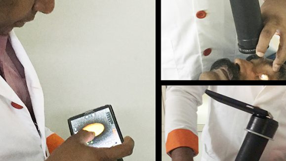
The advent of fundus imaging makes it possible for ophthalmologists and healthcare providers to quickly and safely analyze the retina and diagnose disease. Cost-effective retinal imaging devices are a trend these days, but most of the time, the image quality tends to be substandard as they are often acquired under different lighting environments, using different cameras, and by operators with varying levels of experience. Low-quality fundus images increase uncertainty in clinical observation and may lead to the risk of misdiagnosis.1
Results of a screening study including 5,575 patients showed that about 12% of fundus images are not of adequate quality to be readable by the ophthalmologists.2 Some of the main reasons for low quality in retinal fundus images include uneven illumination, blurring and artifacts, which not only prevent reliable diagnosis by ophthalmologists, but also disturb the performance of automated image analyzing systems.3
Problems with Poor-quality Fundus Images
Advances in artificial intelligence (AI) and deep learning techniques have greatly improved general image enhancement outcomes. Nevertheless, due to the structure of the retina and the unique characteristics of the ophthalmoscope imaging process, natural image enhancement methods cannot be used directly to address low-quality issues in fundus images.
One of the reasons is due to the structure of the retina, which makes it unable to be illuminated internally, hence both incident and reflected imaging beams have to traverse the pupil. Moreover, the spherical geometry of the eye can cause significant inter-reflection, resulting in shading artifacts.4
Anatomical retinal structures are important for clinical diagnosis and should be enhanced in the correcting process.5 And pathological characteristics like hemorrhages, microaneurysms and drusen are usually few pixels wide and appear in circular shapes, which cause them to be easily confused as artifacts and noises.5 Hence, it is important to come out with a fundus image correction method that addresses these challenges that general enhancement techniques do not satisfy.
The Clinical-oriented Fundus Enhancement Network “cofe-Net”
To address the above issues, researchers from China designed a fundus degradation model based on the retinal ophthalmoscope imaging system to simulate low-quality fundus images.5 The model approximates major factors of low-quality fundus images, such as light transmission disturbance, imaging blurring and retinal artifacts.
They then developed a clinical-oriented fundus enhancement network (cofe-NET) to correct the low-quality fundus images for clinical observation and analysis. The cofe-NET suppresses the undesired artifacts based on a low-quality activation (LQA) module, while preserving anatomical retinal structures of fundus images (such as vessel and optic disc regions) by using the retinal structure activation (RSA) module. Pathological characteristics of the fundus images are preserved as well for clinical observation and analysis.
With this, the researchers were able to demonstrate that fundus correction can boost the performances of clinical analysis systems, such as vessel segmentation and disc/cup detection, on poor quality images. Experimental results on both synthetic and real fundus images showed that their algorithm effectively corrects low-quality fundus images without losing retinal details.
The authors were confident that their fundus correction method could assist ophthalmologists in ocular disease diagnosis through retinal fundus image observation and analysis, and be beneficial to medical image applications, such as retinal vessel segmentation and optic disc/cup detection.
References
- Shen Z, Fu H, Shen J, Shao L. Modeling and Enhancing Low-Quality Retinal Fundus Images. IEEE Trans Med Imaging. 2021;40(3):996-1006.
- Philip S, Cowie L M, Olson J A. The impact of the health technology board for Scotland’s grading model on referrals to ophthalmology services. Br J Ophthalmol. 2005; 89(7): 891–896.
- Fu H, Wang B, Shen J, Cui S, Xu Y, Liu J, Shao L. Evaluation of Retinal Image Quality Assessment Networks in Different ColorSpaces. In book: Medical Image Computing and Computer Assisted Intervention – MICCAI 2019, 22nd International Conference, Shenzhen, China, October 13–17, 2019, Proceedings, Part I (pp.48-56)
- Foracchia M, Grisan E, Ruggeri A. Luminosity and contrast normalization in retinal images. Med Image Anal. 2005;9(3):179–190.
- Shen Z, Fu J, Shen J, Shao L. Understanding and Correcting Low-quality Retinal Fundus Images for Clinical Analysis. Available at: https://www.researchgate.net/publication/341342055_Understanding_and_Correcting_Low-quality_Retinal_Fundus_Images_for_Clinical_Analysis Accessed on 14 February 2022.