There is adequate evidence to support the use of intravitreal dexamethasone in diabetic macular edema (DME), according to an expert panel on DME management in the Asian population.
The findings were presented during Saturday’s session on Rolling Out Updates in Retina, moderated by vitreoretinal surgeon Dr. Kenneth Fong, from OasisEye Specialists in Kuala Lumpur, Malaysia.
The right therapy for the right patients
An expert panel of 12 retinal specialists from Singapore, Malaysia, the Philippines, India and Vietnam found that dexamethasone treatment particularly applies to patients who are pseudophakic; those who are anticipated to have poor compliance in follow-up; patients who have been previously vitrectomized; or who have undergone cataract surgery, said Dr. Augustinus Laude, from National Healthcare Group Eye Institute in Singapore.
Intravitreal (IVT) dexamethasone implants are effective in treating persistent DME, both in vitrectomized and non-vitrectomized eyes, and vitrectomy does not influence the efficacy and safety profile of IVT dexamethasone.
He advised that patients who receive IVT dexamethasone should be informed and counseled about possible cataract formation, which typically will become evident about one to two years following treatment.
“Used as adjunctive or combined with intravitreal dexamethasone following cataract surgery is a good option, and helps manage post-surgery recovery quicker, and also minimizes or reduces the risk of postoperative worsening of macular edema in diabetic patients,” he said.
In addition, some patients are non-responders to anti-VEGF, he noted. Switching from anti-VEGF to dexamethasone has been shown to provide better improvement compared to switching within anti-VEGF class.
Next generation iOCT
The next generation EnFocus (Leica, Wetzlar, Germany) intraoperative optical coherence tomography (iOCT) helps surgeons by providing better precision and speed during surgeries, said Dr. Barbara Parolini from Eyecare Clinic, Bresica, Italy, during her presentation on The beauty and usefulness of imaging with intraoperative OCT.
“With the Leica system, the surgeon has more freedom to use it with the foot pedal, and the scans are long with different shapes and sizes, so you can play with your notes,” she said.
EnFocus iOCT provides greater insight during eye surgeries, allowing surgeons to see what lies underneath the surface. This helps surgeons overcome uncertainties during eye surgeries in order to achieve the best possible patient outcome.
EnFocus iOCT can also help answer questions, such as: Is there residual subretinal fluid? Is the glaucoma drainage device in the correct position? Is the corneal graft in the correct orientation?
Further, one of the greatest challenges in vitreoretinal surgery is how to judge residual retinal traction. The EnFocus helps ophthalmic surgeons focus on perfection during surgery, with better visibility compared to the microscope.
“The advantages of the EnFocus iOCT is confirmation of what we see with the microscope, as well as the ability to see what we do not see with the microscope,” she added. “It can really change the strategy of your surgery and improve your surgery.”
Bring on the AI
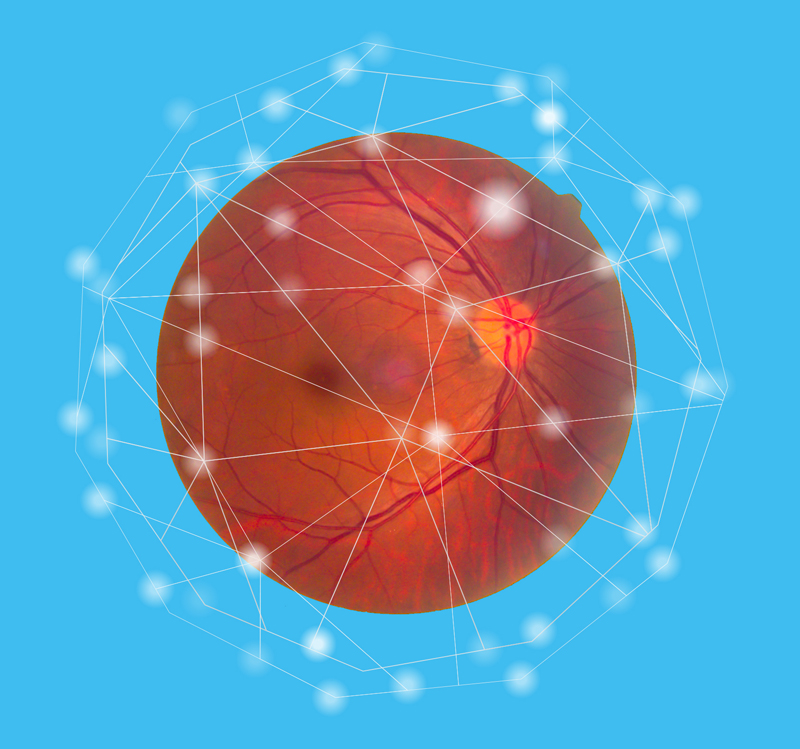
More algorithms for detecting uncommon retinal diseases are in the works as artificial intelligence (AI) applications are increasingly being used in ophthalmology — especially at a time when face-to-face consultations may be limited.
Models for predicting diabetic retinopathy (DR) progression to increase screening intervals are important in the post COVID-19 era, said Dr. Paisan Ruamviboonsuk from the Rajavithi Hospital in Bangkok, Thailand, during his talk on Updates in artificial intelligence for retinal diseases.
Currently, the use of AI primarily concentrates on diseases with a high incidence, such as DR, age-related macular degeneration (AMD), glaucoma, retinopathy of prematurity, age-related or congenital cataract, and retinal vein occlusion.
AI plays a role in common retinal diseases as a screening tool for DR or AMD in populations at risk, as an assisting tool for DME and wet AMD detection, as well as monitoring and decision-making in clinics and research.
For AMD, OCT images still play a major role in AI with the aim of early detection of wet AMD conversion or monitoring treatment.
“More algorithms on detecting other relatively uncommon retinal diseases are on the way,” concluded Dr. Ruamviboonsuk.
Editor’s Note: A version of this article was first published in Issue 3 of CAKE & PIE POST, C&PE 2021 Edition. The CAKE & PIE Expo 2021 was LIVE on June 18-19. All sessions are available on demand until July 19 at expo.mediamice.com upon login.


