
The Zilia Ocular camera’s entry into Canada marks a milestone for oxygen saturation as a biomarker.
Retinal oximetry is officially coming to Canada.
Zilia (Quebec, Canada) announced Thursday that its Ocular retinal camera has achieved Canadian regulatory approval. The nod positions retinal oximetry technology and its implications in oculomics for potential widespread clinical adoption.
“This represents a true paradigm shift,” said Dr. Patrick Sauvageau, CEO and co-founder of Zilia. “By measuring retinal oxygenation in real time, we aim to help clinicians detect and manage eye diseases such as glaucoma, diabetic retinopathy and age-related macular degeneration more effectively.”
READ MORE: Seeing the Unseen: Adaptive Optics Enters the Clinical Arena at ARVO 2025
According to Zilia, Health Canada’s approval of the device marks the first time any regulatory body has authorized a retinal camera to measure oxygen saturation in ocular blood vessels. Other approved ocular oximeters include the CE-marked Oxymap T1 (Oxymap; Reykjavik, Iceland) and the Imedos Dynamic Vessel Analyzer (Imedos Health GmbH; Jena, Germany).
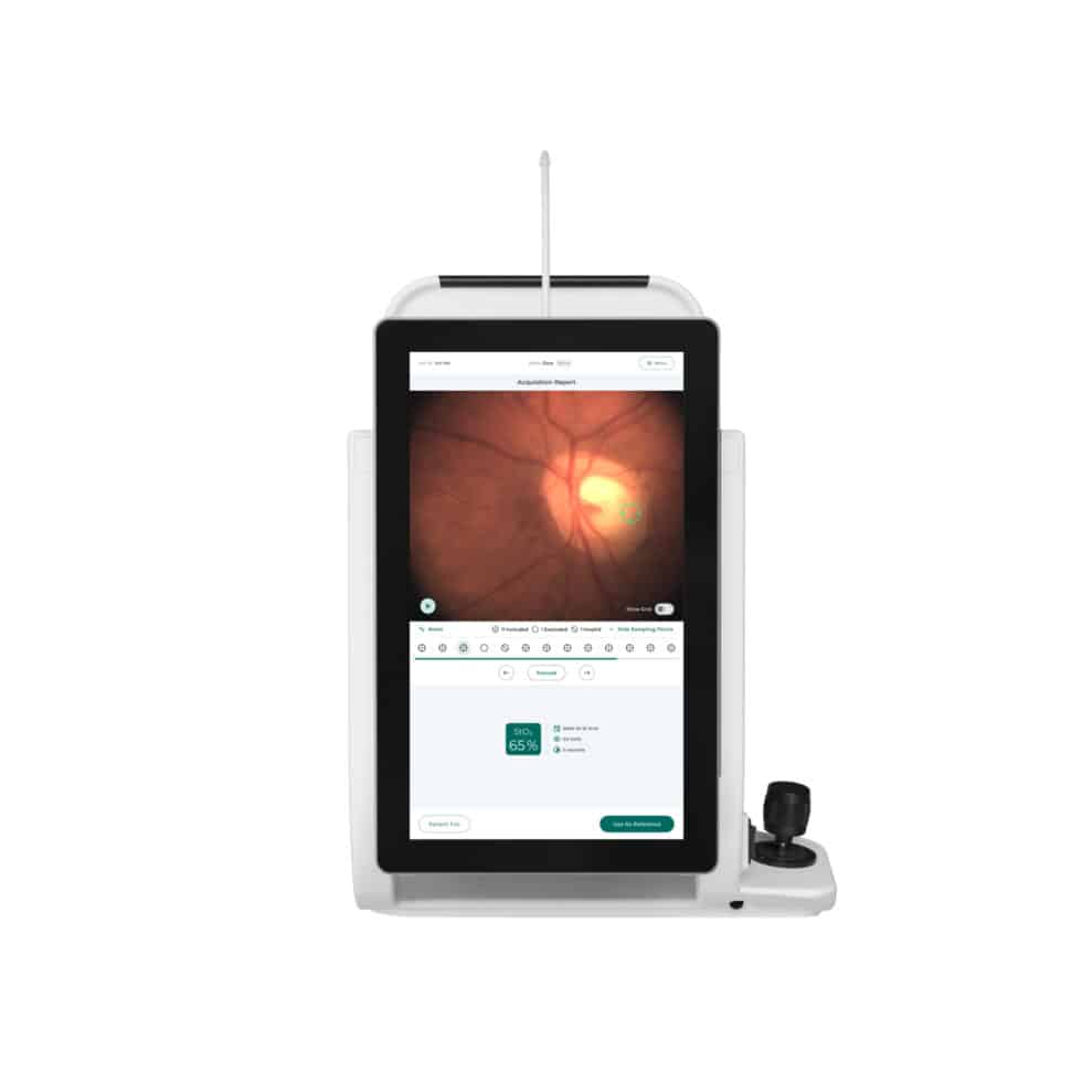
The Zilia Ocular device itself combines advanced photonics with artificial intelligence to provide non-invasive retinal oxygenation measurements. The company plans to roll out the device in Canada in the coming months, with expansion into the United States and Europe forthcoming.
What is retinal oximetry?
Retinal oximetry measures oxygen saturation levels in retinal arteries and veins, providing insights into both ocular and systemic health. The technology works by analyzing how blood absorbs different wavelengths of light, revealing oxygen levels in real time.
Research shows that diabetic retinopathy patients have altered oxygen saturation patterns compared to healthy individuals. These changes often correlate with disease severity and can respond to treatments including anti-vascular endothelial growth factor (anti-VEGF) therapy and laser photocoagulation.1
READ MORE: FDA Approves Roche’s Susvimo for Diabetic Retinopathy
Glaucoma applications have revealed relationships between oxygen saturation patterns and disease progression. The literature suggests that oxygen measurements could provide objective biomarkers for monitoring treatment effectiveness and disease advancement.2
Retinal vascular occlusions also present distinct oximetry signatures, with blocked vessels showing different oxygen patterns than healthy eyes. These measurements could help clinicians monitor treatment progress and assess recovery.3
READ MORE: Visualizing the Retinal Vasculature
Oculomics integration promises systemic health insights
And it’s not just retinal oximetry’s predictive power inside the eye that has people talking.
Retinal oximetry’s integration with oculomics represents a convergence of ocular imaging and systemic health assessment. The retina’s unique properties as the only location where blood vessels can be directly visualized non-invasively make it an optimal window for metabolic monitoring.
Emerging applications could extend beyond ophthalmology to cardiovascular and neurological disease detection. Research suggests potential correlations between retinal oxygen patterns and cardiac health, while studies in neurodegenerative diseases show retinal changes that might precede clinical symptoms.
Editor’s Note: This content is intended exclusively for healthcare professionals. It is not intended for the general public. Products or therapies discussed may not be registered or approved in all jurisdictions, including Singapore.
References
- Blindbæk SL, Peto T, Grauslund J. Correlation between Diabetic Retinopathy Severity and Oxygen Metabolism in Patients with Diabetic Macular Edema during Treatment with Intravitreal Aflibercept. Ophthalmic Res. 2020;63(2):106-113.
- Shughoury A, Mathew S, Arciero J, et al. Retinal oximetry in glaucoma: Investigations and findings reviewed. Acta Ophthalmol. 2020;98(6):559-571.
- Belamkar AV, Jabbehdari S, Harris A, et al. Clinical implications of retinal oximetry in retinal vein occlusion: A review. Acta Ophthalmol. 2022;100(6):624-631.


