AMD gets an upgrade as experts decode the future of imaging, treatments and take-home tech.
Leading retina specialists gathered on the third day of the 40th Congress of the Asia-Pacific Academy of Ophthalmology (APAO 2025)—held in conjunction with the 83rd Annual Conference of the All India Ophthalmological Society (AIOC 2025)—to share the latest developments in age-related macular degeneration (AMD) management and related diseases. The symposium covered advances in imaging, home monitoring, polypoidal choroidal vasculopathy (PCV) and more.
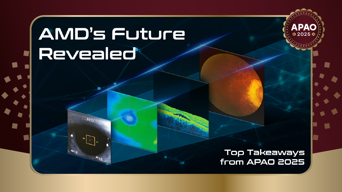
A new imaging era for AMD
Dr. Srinivas Sadda (USA) believes retinal imaging technologies have evolved—and the new guard is leading to better visualizations and measurements of geographic atrophy (GA).
While flash color photography was historically the gold standard, newer modalities now offer superior visualization. “Atrophy can be assessed and measured with multimodal imaging,” noted Dr. Sadda. “I think color photography, or flash color, is the past. The present has been blue autofluorescence, but for my money, it’s these newer technologies that we’re going to be using going forward.”
Dr. Sadda also acknowledged the advantages that optical coherence tomography (OCT) and optical coherence tomography angiography (OCT-A) have displayed supplanted dye-based angiography for routine management—though indocyanine green angiography (ICG) remains the gold standard for PCV diagnosis. He also discussed the importance of recognizing different types of neovascularization using modern tools, as they have distinct prognostic implications.
The promise and limitations of GA treatments
Dr. Neil Bressler (USA) provided a balanced assessment of the recently approved treatments for geographic atrophy—one of the dominant debates in retina over the past year. While there’s excitement about having FDA-approved options, he outlined several key limitations of these newly approved complement inhibitors.
While these treatments slow GA growth by about 20% relative to placebo, said Dr. Bressler, the absolute difference is minimal—only about 0.4mm² at one year. “The magnitude of the anatomic differences is very small,” he explained. “You can barely see it.”
More concerning, these anatomic benefits haven’t translated to functional improvements. “The anatomic benefits were not associated with any clinically meaningful benefit across visual acuity, low luminance or reading speed,” Dr. Bressler stated. “With most macular diseases, including geographic atrophy, vision outcomes should trump anatomic outcomes.”
Safety concerns include increased risk of neovascular AMD and rare but serious cases of occlusive retinal vasculitis. As a result, the European Medicines Agency (EMA) has repeatedly refused marketing authorization, citing unfavorable risk-benefit profiles—though both Izervay (Astellas Pharma; Tokyo, Japan) and Syfovre (Apellis; Waltham, Massachusetts, USA) are approved for use in the United States.
Limited vitamin supplementation evidence for GA
In a separate presentation, Dr. Bressler addressed vitamin supplementation for GA and retinitis pigmentosa (RP). While the AREDS and AREDS2 supplements reduce the risk of progression to neovascular AMD, evidence does not support benefits for GA development or progression.
“Despite the ease of accessing vitamin A, and despite the potentially devastating effects of GA from AMD, or from RP, vitamin supplementation for either of these retinal diseases may not be warranted at this time,” Dr. Bressler said.
The potential and pitfalls of home monitoring
Is OCT monitoring coming home in 2025?
It might be, but that doesn’t mean caution should be abandoned, argued Assoc. Prof. Gavin Tan (Singapore) as he discussed the evolving landscape of home monitoring for AMD.
As treatment intervals extend in exudative retinal disease, the hope is that home OCT can mitigate the increased risk of missing disease activity between clinic visits. But there are also drawbacks.
“Home monitoring holds potential,” said Dr. Tan. However, he also cautioned that “hyperacuity-based tests have limitations in accuracy” and “may perform worse in real-world studies.”
While specialized devices like the ForeSeeHome monitoring device (Notal Vision; Virginia, USA) have shown efficacy in clinical trials, smartphone-based applications have demonstrated poor diagnostic accuracy in real-world settings. Patient compliance also remains a significant challenge, with usage declining over time.
Besides this, the practical realities of the device also put a damper on things—for now, at least. “Logistics, costs and compliance issues need to be addressed” before widespread adoption, Dr. Tan said.
Pachychoroid: Defining a complex condition
Dr. Xinyuan Zhang (China) discussed recent advances made by her team in understanding pachychoroid disease (PCD). Diagnostic criteria remain poorly defined, and her lab has developed objective criteria integrating demographic and clinical factors including age, sex and refraction.
“Thick choroids can also exist in healthy individuals, requiring comprehensive imaging evidence for PCD diagnosis,” Dr. Zhang explained. Her research has focused on the role of lipid metabolism in pachychoroid pathogenesis, with AI-based tools showing potential for improving diagnosis.
Persistent challenges in PCV management
Assoc. Prof. Voraporn Chaikitmongkol (Thailand) outlined ongoing challenges in managing PCV, despite therapeutic advances. While diagnosis has improved, treatment selection and long-term management remain difficult.
“PCV is a disease with a highly recurrent nature, where long-term recurring fluid or massive hemorrhage could occur,” Dr. Chaikitmongkol cautioned.
She presented case examples demonstrating variable responses to anti-VEGF monotherapy, with some patients requiring switching between agents or rescue photodynamic therapy (PDT). Complete polypoidal regression, a key predictor of reduced recurrence risk, is achieved in only 42% to 51% of patients with anti-VEGF monotherapy, compared to 80% with PDT.
But it’s not always just about the medical side of things, Dr. Chaikitmongkol argued, pointing to another area where disease outcomes can be improved. “Patient education is important to help better understand the chronicity and importance of follow-up,” she said.
Novel functional testing shows promise
Dr. Mark Pennesi (USA) presented encouraging data on split-spectrum amplitude decorrelation optoretinography (SSADOR), a new technique for objectively measuring photoreceptor function.
“This delivers an objective and reliable measure of photoreceptor function in both normal and patients with IRDs,” Dr. Pennesi explained. The technology correlates well with microperimetry in rod-cone dystrophies and may detect early cone dysfunction before other tests.
Over the course of the talk, Dr. Pennesi balanced both progress and persistent challenges in AMD management. While new imaging techniques, treatments and monitoring approaches show promise, questions remain about optimal patient selection, real-world effectiveness and long-term outcomes.
Editor’s Note: Reporting for this story took place during the 40th Congress of the Asia-Pacific Academy of Ophthalmology (APAO 2025), being held in conjunction with the 83rd Annual Conference of the All India Ophthalmological Society (AIOC 2025) from 3-6 April in New Delhi, India.


