![[Heidelberg] Website article thumbnail 01](https://piemagazine.org/wp-content/uploads/sites/4/2025/03/Heidelberg-Website-article-thumbnail-01.png)
As recommended by the Clinical Director at Heidelberg Engineering, Christopher Mody
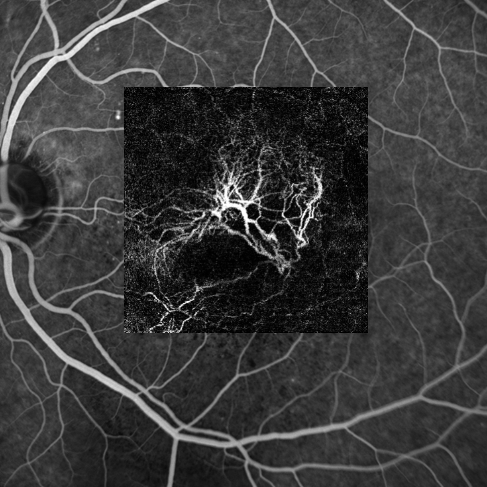
In vivo fluorescein angiography (FA), first described in 1961, has enabled the visualization of the retinal vasculature for over sixty years. High-quality video recordings of vascular flow can be captured using confocal scanning laser ophthalmoscopy (cSLO). This dynamic component of angiographic imaging adds an extra dimension to image interpretation and diagnosis. FA provides a clear visualization of the foveal avascular zone (FAZ) and the outline of the foveal arcade, offering clinicians valuable insights into a patient’s visual performance and potential. Identifying these features is an essential aspect of FA studies. However, a limitation of FA is that, as a two-dimensional projection of a three-dimensional vascular structure, it does not allow for the differentiation of the distinct capillary plexuses within the retina.
OCTA and the retinal structures
Optical coherence tomography (OCT) has revolutionized imaging of retinal structures, greatly enhancing our understanding of chorioretinal pathology in vivo. OCT angiography (OCTA) enables non-invasive imaging of blood flow and provides high-resolution transverse section (en-face) images of retinal vascular patterns. The fundamental principle behind OCTA is that, in a static eye, the only moving structures are the blood cells flowing within the vessels. OCTA can differentiate moving cells from static retinal tissue, detecting flow signals through consecutive B-Scans recorded at the same retinal location, which are then displayed as transverse section images.
OCTA and motion artefacts
However, OCTA images are susceptible to motion artefacts. The SPECTRALIS diagnostic imaging platform from Heidelberg Engineering addresses this issue by employing a live eye tracking system, allowing for precise follow-up scans and minimizing motion-related distortions. The retina contains four distinct vascular plexuses, each with unique morphology tailored to meet the metabolic demands of specific retinal tissues. The average diameter of a retinal capillary, including the lumen, is approximately 8 microns, with blood flow measuring around 6.4 microns in diameter. To accurately capture capillary flow and blood vessel patterns, an OCTA system must meet or exceed this resolution. The SPECTRALIS OCT Angiography Module achieves a lateral resolution of 5.7 microns and an axial resolution of 3.9 microns per pixel, enabling detailed visualization of all four vascular plexuses. This high resolution ensures diagnostic confidence and clinical certainty.
OCTA and imaging quality
Another critical factor in imaging vascular flow with OCTA is the non-linear nature of erythrocyte movement. Variations in blood cell size, shape and orientation can cause clustering at choke points, particularly at vascular bifurcations or in tortuous vessels. As a result, flow may not be detected at a given moment during sampling. Multi-sampling is essential for detecting this non-linear flow. The SPECTRALIS OCT Angiography Module addresses this challenge through a combination of automatic real-time (ART) multi-sampling and TruTrack dual-beam live eye tracking, which mitigate motion artefacts and generate accurate flow information. This technology produces exceptionally high-quality OCTA images, ensuring consistent and reliable documentation of capillary blood flow over time.
Editor’s Note: A version of this article was first published on PIE magazine Issue 34.


