Research in OCT transitioned in the early 2000s from time domain optical coherence tomography (TD-OCT) to spectral domain OCT (SD-OCT). Now, it is again at a transition point from SD-OCT to swept-source OCT (SS-OCT). Certainly the latest developments in SS-OCT are appreciated. Optical coherence tomography angiography (OCTA) – a very new and exciting development in acquiring ophthalmic images from patients, also adds to the visualization landscape.
“When time domain OCT came out, I was fascinated by the images produced, but when spectral domain OCT started churning out beautiful images, I found it very impressive. Certainly, it helped a lot in our technical diagnosis and decision-making, to make better choices for our patients,” said Dr. Augustinus Laude, adjunct associate professor and senior consultant in the National Healthcare Group Eye Institute (NHGEI) at Tan Tock Seng Hospital Singapore (TTHS).
“The transition from the time domain to the spectral domain OCT was very natural, because the change is very obvious. You can get better quality images. You can see much better resolution and that gives us more information about the patient,” explained Dr. Laude.
However, the main issue is that there are still many uncertainties among today’s OCT options. Part of the reason is because of the variability in the type of images acquired. There are also uncertainties about what is actually being seen, because in OCTA, images are acquired by the movements of the red blood cells in the blood vessels.
“We are aware if the blood vessels travel too fast or too slow, the images may not be acquired. So, if we see an image that is generated then disappears, it may be difficult to interpret whether it is due to lack of flow, or difficulty in image acquisition. So, this will lead to uncertainty about what it is we are seeing. We as humans will try to figure out what happened, and it may not be the accurate answer,” added Dr. Laude.
OCTA brings a host of advantages over traditional angiography. Firstly, the time taken for OCTA image acquisition can be completed in a much shorter time. Secondly, it is a much safer and non-invasive procedure that does not involve injecting any medications into the patient. To be able to acquire the images in the shortest time and in the safest way possible is a very good advantage. On the other hand, doctors have to interpret what is shown in the images and decide whether or not it is the pathology that they think they are seeing. Traditional invasive OCTs are still needed in certain circumstances, but doctors are doing less and less of them.
“In the vast majority of cases, such as in patients with macular degeneration, if there are signs of macular degeneration in the OCT, I can get an OCTA of the blood vessels, and that is enough for me to be comfortable making a diagnosis,” said Dr. Nadia Waheed, retinal specialist at the New England Eye Center and director of the Boston Image Reading Center. “Similarly, I use it to visualize the vascular changes that happen in diabetics. If I have a proliferative diabetic retinopathy patient, I’ll often use the OCTA over the area of the abnormal vascular degeneration to make sure that it is truly proliferative and not just due to vascular abnormalities. Normally, I will get my answer through an OCTA. If I don’t, I will go to the next step, which is to do fluorescence or an ICG.”
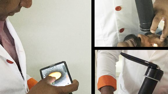
Now, instead of just looking at the surface, we can look into the retina and into the underlying tissues. That is the excitement with new technologies such as the Triton swept source OCT (Topcon, Tokyo, Japan).
“It is one of the first instruments that besides having the ability to take fundus photographs, auto-fluorescence and standard fluorescent angiograms, it also comes with swept source OCT, and it will eventually have the ability to perform angio-OCTs as well,” said Dr. Victor Gonzales, retinal specialist and medical director of the Valley Retina Institute, as well as head of Retinal Research Center, Valley Retina Research (Texas, USA). “It’s an all-inclusive machine that saves space. You don’t need multiple machines to do different things.”
Artificial intelligence (AI) seems to be the buzzword today, and there are many applications that are being explored. It is a very powerful tool, and it is possible to find applications for it within the field of OCTA as well.
“One of the potential applications of AI would be to train an algorithm to be able to rapidly classify the acquired images into either disease or non-disease state, and further into more categories as required,” said Dr. Laude.
It is advantageous to use deep learning particularly to process vast amounts of information which is very time consuming and requires substantial manpower to look through and process manually. AI can be used to help predict who is going to have a disease as well.
“AI makes it possible to have a very precise characterization of the retina by analyzing OCT scans. It is accurate in identifying lesions – it not only makes it possible to identify lesions, but to quantify it in a precise manner,” said Dr. Hrvoje Bogunovic, Department of Ophthalmology, Medical University of Vienna (Vienna, Austria).
“Before the advent of AI, this was not possible because clinicians did not have time to view every single slide of the OCT to make annotations. At this moment, this type of measurement simply does not exist. So, this will bring the whole practice to a new level, and the best part is we already have the technology ready to be used,” added Dr. Bogunovic.
Undoubtedly, many new discoveries and developments in research and development are underway with regard to OCT, OCTA, SS-OCTA and AI. We look forward to more exciting developments and advances in treatments to help patients improve their vision and quality of life in the future.
Editor’s Note: This article is an excerpt from PIE Talks 2: Interviews at ARVO 2018 Honolulu. Search YouTube for “PIE Talks Episode 02” for the complete videos of the individual interviews.
“When time domain OCT came out, I was fascinated by the images produced, but when spectral domain OCT started churning out beautiful images, I found it very impressive. Certainly, it helped a lot in our technical diagnosis and decision-making, to make better choices for our patients.”
– Dr. Augustinus Laude, Senior Consultant, National Healthcare Group Eye Institute, Tan Tock Seng Hospital Singapore
“AI makes it possible to have a very precise characterization of the retina by analyzing OCT scans. It is accurate in identifying lesions – it not only makes it possible to identify lesions, but to quantify it in a precise, manner.”
– Dr. Hrvoje Bogunovic, Department of Ophthalmology, Medical University of Vienna (Vienna, Austria)