This dry AMD session outlined hidden risks, trial bottlenecks and the growing promise of precision.
On Day 1 of the 25th Congress of the European Society of Retina Specialists (EURETINA 2025), the Nonexudative AMD symposium gathered five international experts who examined supplements, imaging biomarkers, structure-function mapping, AI models and the evolving treatment pipeline in meticulous detail.
A unifying current ran through the talks, framing geographic atrophy (GA) as layered, multifactorial and anything but uniform, demanding sharper endpoints, individualized risk assessment and earlier, smarter intervention.
READ MORE: Advancing the Management of Geographic Atrophy
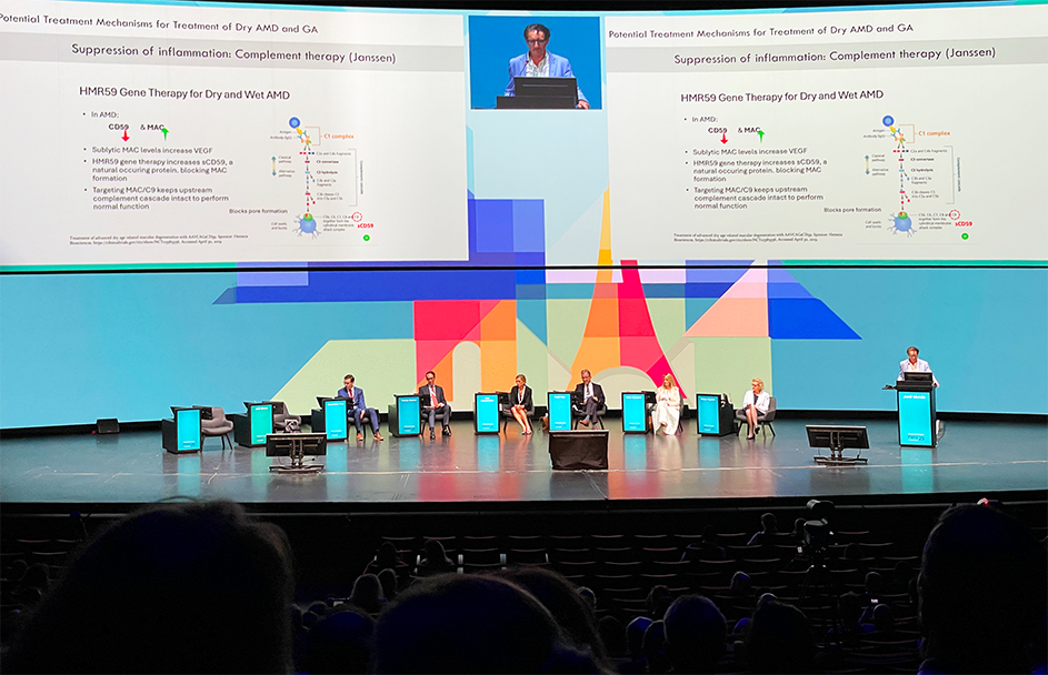
What’s next in the dry AMD pipeline?
Dr. Jordi Monés (Spain) opened the age-related macular degeneration (AMD) session with a sober but forward-looking assessment of where the field stands on GA.
Despite recent approvals of complement inhibitors such as pegcetacoplan (Apellis Pharmaceuticals; Massachusetts, USA) and avacincaptad pegol (Astellas Pharma; Tokyo, Japan), progress in Europe has felt glacial, slowed by regulatory lag and trial design hurdles.
READ MORE: FDA Expands Label for Izervay in Geographic Atrophy Treatment
For Dr. Monés, the central issue is precision. GA is not a monolith but a spectrum of phenotypes that are sometimes fast, slow and resistant, and “lumping all patients into one category,” he warned, risks diluting meaningful results.
He emphasized the importance of phenotypic stratification and argued for incorporating structural biomarkers such as subretinal drusenoid deposits and photoreceptor thinning as enrichment tools and endpoints, especially in earlier pre-atrophic stages where intervention may be most effective. Adaptive trial designs that capture both structure and function will be key to unlocking progress.
“The disease is so different and the phenotypes are so different,” Dr. Monés said. “That is like if you were asking, ‘Are you going to treat skin cancer and pancreatic cancer with the same drug?’ No way, it’s probably a very different animal. So we will need to do different therapies to different patients and maybe combined therapies.”
The future, he concluded, lies in tailored, earlier intervention that shifts GA from “wait and watch” to “diagnose and intervene.”
Do supplements hold back GA?
Dr. Tiarnan Keenan (USA) revisited the AREDS story with a sharper lens, asking whether supplements might still have a role in GA. While past analyses suggested little impact on lesion growth, his re-examination of AREDS and AREDS2 told a more nuanced tale.
By applying a “growth-to-center” metric that tracked how quickly lesions encroach on the fovea, Dr. Keenan found signals that standard area-based measures had missed. Supplementation slowed GA’s advance toward central vision by roughly 35%, and when lutein and zeaxanthin were factored in, the risk of foveal involvement was cut by more than half.1
These effects were most apparent in extrafoveal lesions, where functional vision remains intact. “The area metric is blunt,” he noted. “It misses the functional relevance of where the lesion is progressing.”
Zinc and other components showed subtler, stage-dependent patterns, though none were definitive. Importantly, these insights emerged from secondary analyses, so confounding remains a concern.
“We need a new dedicated prospective trial looking specifically for this as part of the AREDS 3 program… even when people develop geographic atrophy and particularly in that extrafoveal case,” said Dr. Keenan, stressing the need for validation.
For now, supplements remain safe, inexpensive and accessible, and provide a modest but meaningful tool in an otherwise limited armamentarium.
READ MORE: Inflammasome Therapeutics Reports 3-Month Trial Data for Inflammasome Inhibitor in GA
Smarter endpoints for trials
“Best-corrected visual acuity alone is no longer enough.” With that, Dr. Stela Vujosevic (Italy) set the tone for her call to rethink endpoints in nonexudative AMD.
While OCT holds the fort as the backbone of diagnosis, she stressed that structure must be paired with function if trials are to reflect real-world patient experience. “Structural imaging tells us what’s there,” she explained, “but functional tests tell us what the patient experiences.”
Dr. Vujosevic highlighted tools such as low-luminance visual acuity (LLVA), microperimetry, dark adaptometry and contrast sensitivity that are all validated in longitudinal studies like MACUSTAR. Their utility varies by disease stage: extrafoveal lesions align best with microperimetry and LLVA, while fovea-involving disease may still justify BCVA.
Importantly, evidence suggests functional decline often precedes structural change, meaning these assessments can flag vision loss earlier than imaging alone.
READ MORE: Retinal Imaging and AI-Powered Analysis in the Palm of Your Hand
Dr. Vujosevic also pointed to OAKS trial data showing that microperimetry demonstrated preserved function with pegcetacoplan, illustrating how smarter endpoints can capture therapeutic benefits that BCVA misses. She concluded that “the composite endpoints of structure and function that are tailored to GA progression, patient needs would be ideal for upcoming clinical trials, allowing earlier intervention”.
For physicians, this means moving beyond lesion size to patient-centered measures that better guide care.
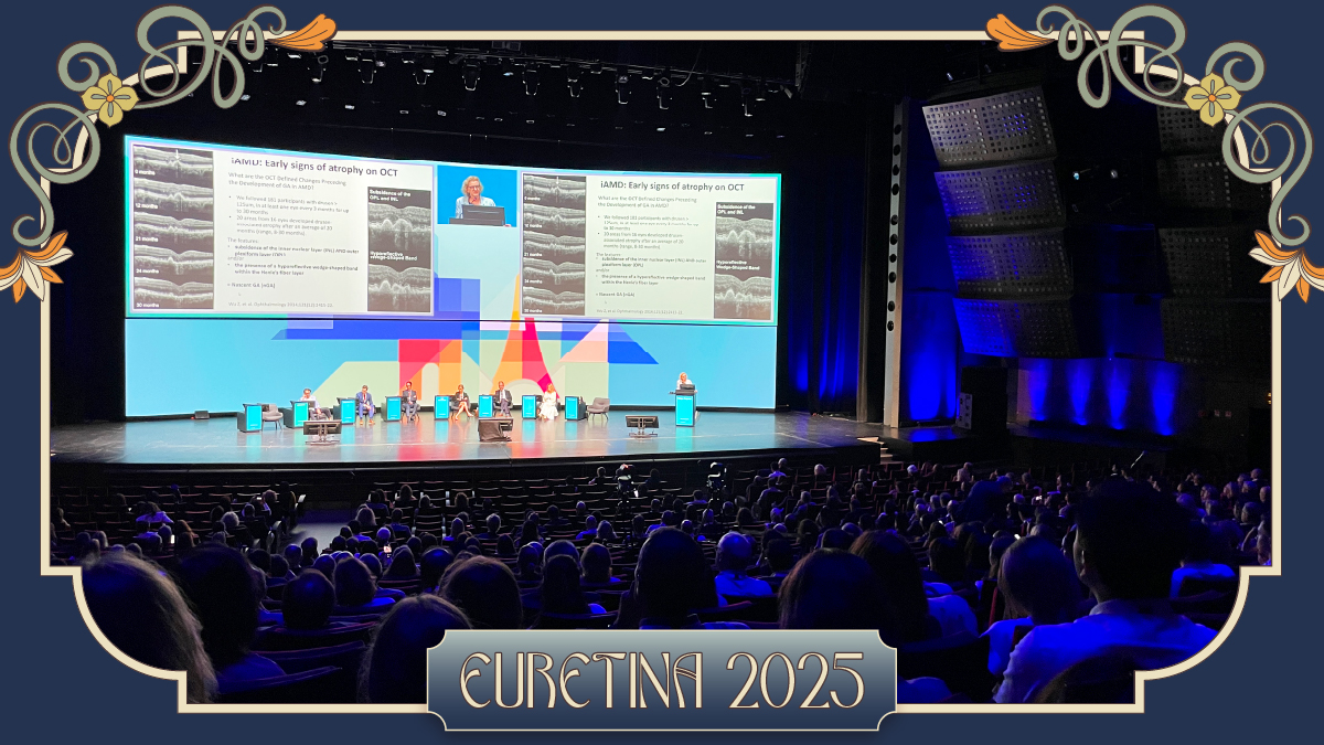
Reading the signs of risk
Prof. Robyn Guymer (Australia) shifted the spotlight to intermediate AMD—the gray zone where disease is present but its future course is unpredictable.
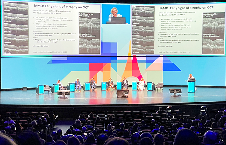
She argued that fundus-based classifications undersell the precision now possible with OCT, where biomarkers such as nascent GA, incomplete retinal pigment epithelium (RPE) and outer retinal atrophy (iRORA), and complete RPE and outer retinal atrophy (cRORA) are rewriting the risk playbook.
Hazard ratios climb as high as 78 for nascent GA, a stark measure of their predictive weight. Retinal layer changes, including early loss of the ellipsoid zone and external limiting membrane, can also flag progression years before overt GA emerges.
Subretinal drusenoid deposits (SDDs) drew particular focus. “SDDs are not just a variant,” Prof. Guymer noted. “They represent a fundamentally different phenotype with faster progression and poorer outcomes.”
Differentiating SDDs from softer drusen is critical, not only for prognosis but also for eventual treatment matching. Her group has even released an AI-driven algorithm to standardize SDD detection across large datasets, an advance that could sharpen enrichment strategies in future trials.
“If you want to be highly specific in your predicting ability, then we’re really not very accurate. We’re less than 50% likely to predict who’s most likely to progress,” said Prof. Guymer, acknowledging current limitations.
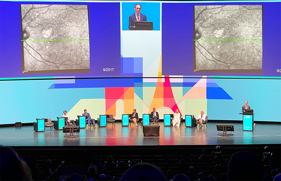
Predicting outcomes in three dimensions
Closing the session, Dr. Maximilian Pfau (Germany) reframed GA through a three-dimensional lens.
“It’s not a two-dimensional disease,” he declared, introducing the concept of GA as a funnel of photoreceptor degeneration extending far beyond the visible atrophic border. Using AI-powered OCT segmentation and deviation maps, his team tracked outer nuclear layer (ONL) thinning, a marker that predicts both future lesion growth and therapeutic response.
Patients with broader ONL loss at baseline progressed more quickly, yet paradoxically responded better to treatment. This convergence of risk and opportunity highlights the potential of phenotype-specific modifiers, such as subretinal drusenoid deposits, to guide therapy allocation and refine trial design.
Dr. Pfau likened this to treatment effect modification, well established in intermediate AMD and now reaching into GA.
Beyond anatomy, his team has built AI models capable of inferring functional vision directly from OCT, effectively bypassing the need for microperimetry. These sensitivity maps offer higher spatial resolution and greater reliability for longitudinal tracking.
As he explained, “AI-based inferred sensitivity can actually be used as a surrogate of microperimetry in real world clinics and it provides much better reliability and spatial resolution.”
For Dr. Pfau, AI is not an accessory but the engine powering smarter endpoints, earlier intervention and more personalized care.
READ MORE: 4 Reasons Why AI Is Important in Medical Image Analysis
Playing the long game
While the silver bullet for GA is yet to be found, Thursday’s session showed how rapidly the landscape is being redrawn. From supplements and OCT biomarkers to functional testing, gene and mitochondrial strategies, and AI models that map disease in three dimensions, the toolbox is expanding.
The experts agreed that without sharper trial designs and better patient segmentation, even the strongest candidates risk falling short. With earlier detection, composite endpoints and precision strategies that match therapy to phenotype, nonexudative AMD may finally move from “untreatable” toward “manageable,” giving patients the real options that they’ve been waiting for.
Follow our daily EURETINA coverage for expert insights and groundbreaking retina updates.
Editor’s Note: The 25th EURETINA Congress is being held from 4-7 September, in Paris, France. Reporting for this story took place during the event. This content is intended exclusively for healthcare professionals. It is not intended for the general public. Products or therapies discussed may not be registered or approved in all jurisdictions, including Singapore.
Reference
- Keenan TDL, Agron E, Keane PA, Domalpally A, Chew EY. Oral antioxidant and lutein/zeaxanthin supplements slow geographic atrophy progression to the fovea in age-related macular degeneration. Ophthalmol. 2025;132(1):14-29.