Advancing Retinal Disease Management Through Multimodal Imaging: Effective retinal disease management requires a multifaceted approach integrating various diagnostic modalities. As outlined previously, combining various small-field/high resolution and ultra-widefield imaging modalities, such as multiwavelength imaging (e.g. red/green or red/green/blue), OCT and OCTA, as well as fundus autofluorescence (FA), fluorescein/indocyanine green (FA/ICG) angiography (FA) comprehensive information can be acquired [1] (see Figures 1-3). However, a holistic diagnostic process also requires additional testing, incorporating both subjective and objective functional assessments.
Functional testing – A Key Management Tool: Functional tests like best-corrected visual acuity (BCVA) and visual field testing remain fundamental. Though subjective, these tests remain indispensable when structural findings don’t align with a patient’s symptoms.
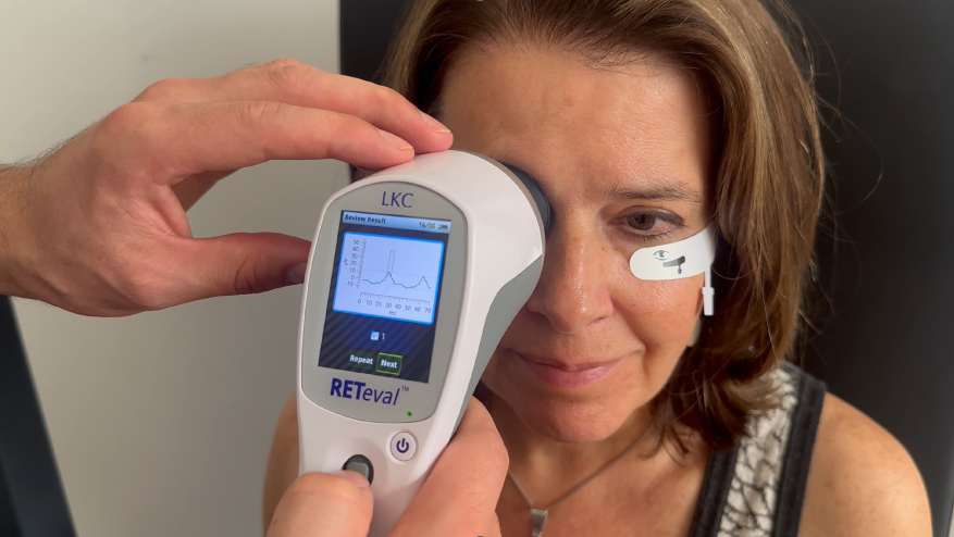
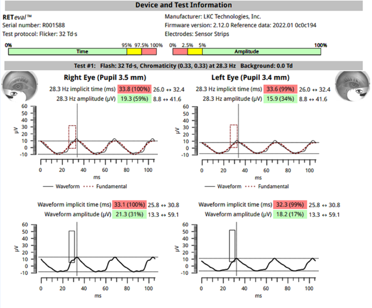
Role of visual electrophysiology as objective functional test: Introduced in the 1940s, visual electrophysiology objectively measures retinal function by detecting electrical responses to light stimuli [2]. Unlike BCVA and visual field testing, it provides objective data and is particularly valuable for diagnosing inherited retinal diseases or cases where subjective tests and structural imaging lack adequate clinical correlation [2]. Although visual electrophysiology is less clinically adopted, studies confirm its role in disease detection [3-5], treatment monitoring [6-8], assessing retinal ischaemia, and progression prediction [9,10] in conditions like diabetic retinopathy, glaucoma or vein occlusion. With modern, user- and patient- friendly ERG devices, like RETeval™ (Figure 4), we notice broader clinical adoption. Having test results on the spot without waiting for information has played a significant role in private practices like ours, where patients come from both close and distant locations.
Combining Functional and Structural Assessments: Functional tests like BCVA and visual field tests reflect a patient’s visual experience, while ERG rather detects disease processes [11]. Structural imaging— small-field/high resolution and ultra-widefield multiwavelength imaging, OCT, OCT-A,FA/ICG angiography and fundus autofluorescence—offer detailed views of retinal anatomy and pathology. Together, these modalities enhance disease staging and treatment planning. For example, in Stargardts, ultra widefield imaging assesses mid and peripheral retinal integrity [12], OCT details macular changes [13], and ERG assesses disease activity across the retina and risk of progression [14]. With more user-friendly ERG devices, these benefits can be extended to acquired retinal diseases such as diabetic retinopathy, or as in a case of ours at The Retina Clinic London, a patient with inflammatory disease, where ERG can be a key biomarker.
“With modern, user and patient- friendly ERG devices, like RETeval (Figure 1), we notice broader clinical adoption. Having test results on spot without waiting for information has played a significant role in private practices like ours, where patients are coming from distant locations.”
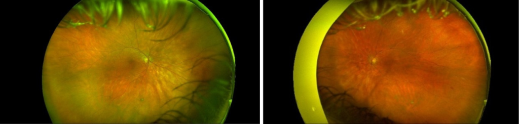
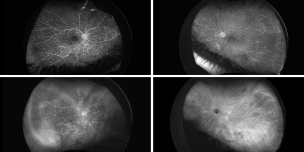
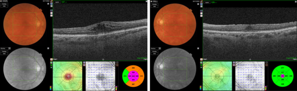
A 62-year-old patient, diagnosed with bilateral intraocular inflammation in Birdshot Chorioretinopathy, suffered from ongoing reduced visual acuity. Initial structural imaging during ongoing treatment (Figure 5-7), including ultra-widefield multiwavelength imaging and OCT, was inconclusive in determining or ruling out the presence of active inflammation. However, a RETeval ERG test showed prolonged implicit times, confirming ongoing inflammatory activity. This objective functional data guided further clinical management decisions, thus highlighting the critical role of integrating functional testing with multimodal imaging in complex cases.
Integrating objective functional assessments like ERG with structural multimodal imaging provides a holistic and therefore beyond comprehensive assessment of disease activity, allowing earlier and more accurate diagnosis and patient management. Functional tests such as BCVA and visual field testing are essential for patient evaluation. However, objective tests like ERG offer deeper insights, often detecting abnormalities earlier than structural imaging alone. In several conditions, ERG predicts disease progression and is highly sensitive to treatment effects. Structural imaging modalities—including small-field/high resolution and imaging such as multiwavelength, OCT, OCT-A, fluorescein/ICG angiography and fundus autofluorescence—of course remain essential.
A multimodal approach combining objective functional and structural testing is sine qua non for optimizing patient outcomes in the management of retinal disease.
References
- Stanga P. RGB: New Hues on the Block. PIE Magazine, Issue 33, January 2025, Page 14-16
- Robson AG, Frishman LJ, Grigg J, et al. ISCEV Standard for full-field clinical electroretinography (2022 update). Doc Ophthalmol. 2022 Jun;144(3):165-177.
- Zeng Y, Cao D, Yang D, et al. Screening for diabetic retinopathy in diabetic patients with a mydriasis-free, full-field flicker electroretinogram recording device. Doc Ophthalmol. 2020;140(3):211-220. [Epub 2019 Nov 12]
- Creel DJ. Electroretinograms. Handb Clin Neurol. 2019;160:481-493.
- Ng JS. The time is now for visual electrophysiology. Int J Ophthalmic Pathol. 2012;1:1
- Birch DG. Surrogate electroretinographic markers for assessing therapeutic efficacy in the retina. Expert Rev Mol Diagn. 2004;4(5):693-703.
- Tang J, Hui F, Hadoux X, Soares B, Jamieson M, van Wijngaarden P, Coote M, Crowston JG. Short-Term Changes in the Photopic Negative Response Following Intraocular Pressure Lowering in Glaucoma. Invest Ophthalmol Vis Sci. 2020;61(10):16.
- Yasuda S, Kachi S, Ueno S, Piao CH, Terasaki H. Flicker electroretinograms before and after intravitreal ranibizumab injection in eyes with central retinal vein occlusion. Acta Ophthalmol. 2015;93(6):e465-8.
- Brigell MG, Chiang B, Maa AY, Davis CQ. Enhancing Risk Assessment in Patients with Diabetic Retinopathy by Combining Measures of Retinal Function and Structure. Transl Vis Sci Technol. 2020;9(9):40.
- Larsson J, Andréasson S. Photopic 30 Hz flicker ERG as a predictor for rubeosis in central retinal vein occlusion. Br J Ophthalmol. 2001;85(6):683-685.
- Kühlewein L, Stingl K. Elektrophysiologie in der Augenheilkunde [Electrophysiology in ophthalmology]. Ophthalmologie. 2024;121(12):1001-1010. [Article in German]
- Kumar V. Insights into autofluorescence patterns in Stargardt macular dystrophy using ultra-wide-field imaging. Graefes Arch Clin Exp Ophthalmol. 2017;255(10):1917-1922.
- Guduru A, Lupidi M, Gupta A, Jalali S, Chhablani J. Comparative analysis of autofluorescence and OCT angiography in Stargardt disease. Br J Ophthalmol. 2018;102(9):1204-1207.
- Sajovic J, Meglič A, Hawlina M, Fakin A. Electroretinography as a Biomarker to Monitor the Progression of Stargardt Disease. Int J Mol Sci. 2022;23(24):16161.