Based on a presentation by Dr. Jennifer Kang-Mieler, Associate Professor of Biomedical Engineering at the Illinois Institute of Technology, Chicago, USA, about Long-Acting Drug Delivery Systems.
“There is an urgent need for delivery system that allows for small drug doses, with constant sustained level of exposure over time,” stated Dr. Jennifer Kang-Mieler during a presentation about long-acting drug delivery systems in the retinal vein occlusion (RVO) session at the recently held 11th Asia-Pacific Vitreo-retina Society Congress (APVRS 2017) in Kuala Lumpur Malaysia.
Although intravitreal injections remain the most efficient method of drug delivery to the posterior segment, they are also [sometimes] associated with potential complications such as endopthalmitis, retinal tears/detachment, cataract formation and – notably – the bolus amount of drug delivered to the eye.
At the moment, US Food and Drug Authority (FDA) approved, corticosteroid-containing drug delivery systems act via a slow process of degradation of their encapsulating polymers. However, due to their unique bio-properties, there are constraints with the use of these systems to deliver anti-VEGF agents. Currently, some of the available systems are:
• Microneedles. Micrometer-sized needles deliver drugs into the supra-choroidal space. They have been assessed for delivery of triamcinolone in phase 1 and 2 clinical trials with encouraging safety and efficacy results in patients with RVO.
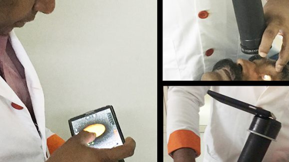
• Microcatheters. Small, illuminated cannulas inserted through sclerotomy which allow for visualization of the supra-choroidal space for accurate drug delivery. These have been evaluated in clinical studies, with no serious intra- or post-operative complications.
• Encapsulated cell technology (ECT). This system consists of cells, genetically modified to produce a specific drug, encapsulated in a semi-permeable hollow fiber membrane. These implants, which are placed in vitreous and can be retrieved, have been shown to continually release drugs for up to 18 months.
• Replenish micropumpsTM. Programmable, refillable, micro-electromechanical systems that deliver continuous or bolus-targeted drugs to the eye up to 12 months.
• The microsphere hydrogel delivery system. Developed by Dr. Kang-Mieler and colleagues, this system contains bio-degradable microspheres with encapsulated anti-VEGF imbedded into a thermos-responsive hydrogel, which confines the microspheres into specific delivery sites. Impressive safety and efficacy results have been shown in non-human primate studies.
Dr. Kang-Mieler concluded that although a variety of approaches to improving drug delivery to the posterior segments are in development, intravitreal injections and/or use of sustained release solid implant intravitreal devices remain the most common techniques at present. Therefore, more innovation is needed as new therapeutic options become available.
Based on a presentation by Dr. Mark Gillies, Professor of Clinical Ophthalmology & Eye Health at the Central Clinical School, University of Sydney, Australia, about Steroids for RVO.
In another presentation, Dr. Mark Gillies provided a historical perspective on the use of steroids in the management of RVO. He noted that the initial clinical development of steroid injections for macular disease was done in Sydney, and earlier studies found little beneficial effect of intravitreal triamcinolone in age-related macular degeneration (AMD). However, further evaluation in patients with diabetic macular edema (DME) showed remarkable results with a doubling of vision gain and reduced macular edema, thereby establishing a role for steroids in the management of DME.
Subsequent clinical studies evaluated the use of steroids in various indications. In the SCORE study, 1 and 4 mgs of intravitreal triamcinolone was compared with standard of care (laser) in patients with RVO. In patients with branch retinal vein occlusion (BRVO), higher rates of cataract and elevated intraocular pressure (IOP) were recorded in the steroid group as compared to laser treatment. However, in those with central retinal vein occlusion (CRVO), the outcomes were better in patients who received triamcinolone, with vision improvements sustained into three years after initiation.
In the GENEVA study, DME study patients received dexamethasone implants (0.7mg, 0.35mg) or sham for RVO. At the 3-month follow-up, there was significant reduction in central macular thickness and improved vision in steroid treated patients as compared to the sham treated. However, at month 6, there was no difference in the outcomes between the group, and further analysis of time-to-first-gain of >15 letters showed better outcomes in the dexamethasone treated patients. The BEVORDEX study provided evidence of optimal dosing interval of steroids in RVO, and concluded that 4-monthly dosing was sufficient to induce sustained reduction in central macular thickness (CMT).
The COMRADE B and C studies compared ranibizumab with dexamethasone treatment in RVO. In these studies, the effects on best corrected visual acuity (BCVA), IOP and CMT with dexamethasone diminished after 2 months, suggesting patients may require repeated dosing before month 6.
The COBALT study evaluated the efficacy and safety of the dexamethasone implant in BRVO-associated macular edema of < 3 months duration. It showed significant mean BCVA improvement with dexamethasone, good IOP control and significant central retinal thickness reduced in patients. Furthermore, majority of the eyes required only one or two injection every 12 months.
Regarding the use of steroids in RVO, Dr. Gillies concluded: “For phakic eyes, use steroids as second line treatment after 6-12 injections of anti-VEGF. However, in pseudophakia, it is best to start with steroids as first line of treatment.”
Based on a presentation by Dr. Raja Narayanan of LV Prasad Eye Institute, India, about Zaltrap and Avastin in RVO: Economy and Effectiveness.
Dr. Raja Narayanan discussed theoretical and practical issues on the use of ziv-aflibercept (ZALTRAP) in the management of RVO.
Aflibercept is a soluble fusion protein of VEGFR1 and VEGFR2 receptors with a strong binding affinity for VEGF-A, VEGF-B and PIGF. And while Ziv-aflibercept received US FDA approval for colorectal cancer, it can also be used off-label for ophthalmological diseases. Ziv-aflibercept and aflibercept are similar – differing mainly in osmolarity (ziv-aflibercept = 1000 mOsm/kg; aflibercept=300 mOsm/kg). This difference is of vital importance as published studies in animals suggest that vitreal osmolarity > 500 mOsm/kg can induce retinal damage. However, given that the vitreous volume in human eyes is three times larger than that in rabbits, an injection of 0.5ml of 1000 mOsm/kg ziv-aflibercept in the human vitreous gets significantly diluted, resulting in a final osmolarity of about 300 mOsm/kg, which is similar to that of aflibercept.
In a recently published a paper, Dr. Narayanan and colleagues showed that safety of intravitreal ziv-aflibercept was acceptable, with no toxicity on ERG, or clinical examination, after a single injection in 12 eyes. To evaluate the efficacy of ZALTRAP in patients with BRVO, Dr. Narayanan and colleagues have injected 88 eyes over the last 2 years, and 42% of these gained more than 15 letters on VA assessment. Although this is an off-label indication, he concluded that ziv-aflibercept may be an option in cases of RVO, at a dose of 1.25mg/0.05ml. “Newer doses of 2mg/0.08ml are under investigation, however larger prospective studies are needed to validate these findings,” said Dr. Narayanan.
Editor’s Note: Search YouTube for “PIE Magazine LIVE @ APVRS” to find the full videos of the talks by Dr. Kang-Mieler, Dr. Gillies, and Dr. Narayanan.