Nearly four percent of children develop poor vision in one eye – known as amblyopia, or more commonly, “lazy eye” – simply because one eye was suppressed or not adequately used during early childhood. This suppression is usually the result of a misalignment of the child’s eyes, such as crossed eyes, or a difference in image quality or focus between the two eyes. Eventually one eye becomes stronger than the other, and over time the weaker eye may become useless. Because amblyopia can be reversed if found and treated early, this is a major public health concern and effective childhood screening needs to take place.
As with most screening programs, finding an assessment that is reliable, cost-effective and that can be performed by non-physician personnel is essential in addressing the large number of individuals who need to be screened. In order to reliably diagnose amblyopia, the detection of reliable central fixation is essential. Yet there remains a need for a commercially available and widely accepted automated screening instrument that can reliably detect strabismus and defocus in young subjects.
The latest contribution on the topic of pediatrics vision screening comes from Dr. Boris Gramatikov, with the Laboratory of Ophthalmic Instrument Development in the Zanvyl Krieger Children’s Eye Center at the Wilmer Institute (Johns Hopkins University School of Medicine), who recently published his work in the journal, BioMedical Engineering OnLine. This laboratory continues to be on the cutting edge of vision screening, with achievements including numerous publications, and four issued or allowed U.S. patents. The work has been supported by an R01 grant from the National Institutes of Health (NIH), an Individual Biomedical Research Award from The Hartwell Foundation, as well as a number of private donors.
Everyone knows that vision assessments in children, like teasing out lack of fixation from simple lack of attention, can be particularly challenging. Dr. Gramatikov’s work discusses how depending on the direction of gaze and the instrument design, the screener produces several signal frequencies that can be utilized in the detection of central fixation. The article also describes how the use of artificial neural networks can allow for a more reliable detection of central fixation and eye alignment, which is essential in diagnosing amblyopia (lazy eye).
To further explaining neural networks, Dr. Gramatikov said: “They are multilayer decision-making mathematical structures that resemble the organization of the neurons in the human brain. They have an input layer that accepts the available data, hidden layers that can be trained (or taught) to represent the interconnections between the neurons at different levels, and an output layer providing the decisions made based on the input data and the network structure and training.”
“This particular network has four inputs, representing normalized spectral powers at four signal frequencies generated by our screening device during retinal birefringence scanning for each eye. The hidden layer contains four neurons. The output suggests presence or absence of central fixation,” he added.
Dr. Gramatikov used the method of backpropagation to train the network, which is a method that calculates the gradient of the loss function with respect to the weights in an artificial neural network. Once the network was trained, validated and tested on a set of controlled calibration data, obtained from 600 measurements from 10 eyes in a previous study, it was tested again on a clinical set of 78 eyes, which were independently diagnosed by an ophthalmologist.
Four inputs for each clinical subject were fed to the artificial neural network, then the output was compared each time with the target, a step in the process which in fact was the doctor’s decision. This allowed for the calculation of the sensitivity and specificity of the artificial neural network when applied to the clinical data.
The results of the tests on the clinical set demonstrated a sensitivity of 0.9851 and specificity of 1.0000, with no false positive decisions and only one false negative decision. Importantly, these results were slightly better than those obtained from the same device and the same signals in their earlier study using statistical methods, including discriminant analysis.
“This work builds upon intensive development work in this field performed in our lab in the past 20 years under the guidance of Dr. David Guyton, a prominent pediatric ophthalmologist, a physicist, and an inventor. Other team members in the past included Drs. David Hunter, Kristi Irsch, Kurt Simons, just to name a few, “shared Dr. Gramatikov.
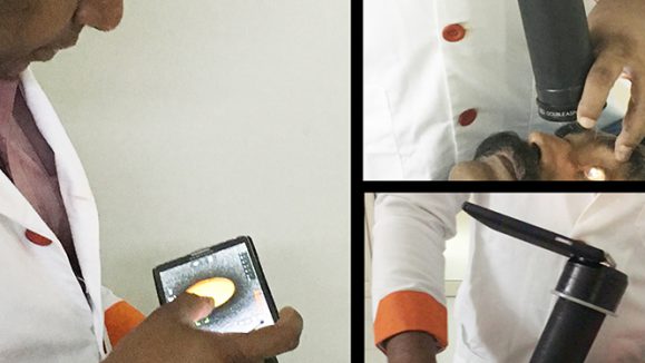
“We have developed an improved pediatric vision screener (PVS) that can reliably detect central fixation, eye alignment and focus. The instrument identifies risk factors for amblyopia, namely eye misalignment and defocus. It uses the birefringence of the human fovea (the most sensitive part of the retina),” he explained.
Birefringence is the property of media (in this case the tiny Henle fibers surrounding the fovea) that change the polarization state of light upon reflection of a beam of polarized light from the fundus in a double pass scanning system. The optics have been reported in more detail in 2014 in the Journal of Biomedical Optics. Dr. Gramatikov notes that circular scanning, as performed by this instrument, generates signals of four distinct frequencies, whose amplitudes are indicative of central fixation or lack thereof.
Although this initial sample size may be considered relatively small, this pediatrics vision screening instrument, in its design and analysis methods, will undoubtedly prove to be incredibly valuable in effectively screening large numbers of children and identifying eye misalignment.
Reference:
Gramatikov BI. Detecting central fixation by means of artificial neural networks in a pediatric vision screener using retinal birefringence scanning. Biomed Eng Online. 2017;16(1):52.