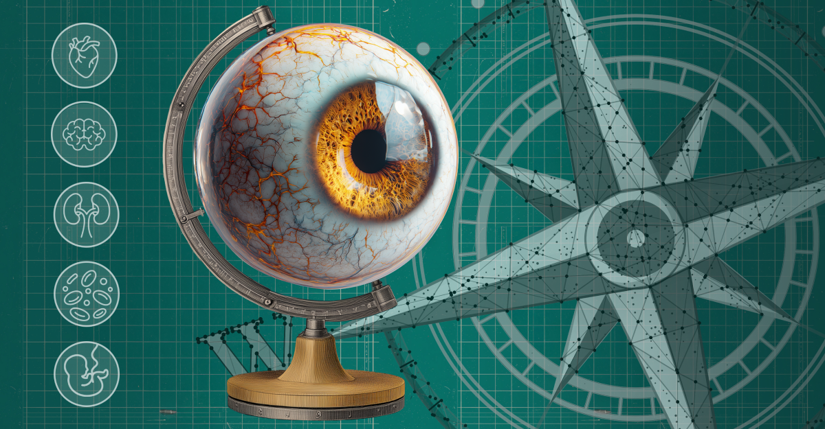
Medicine’s most revealing map is hiding in plain sight. With AI as its compass, the eye is charting systemic disease from the retina outwards, redrawing how clinicians navigate cardiovascular, metabolic and neurologic health long before symptoms appear.
The eye is no longer just the organ of sight, it’s shaping up to be a roadmap of the body itself. Enter oculomics, the science of decoding ocular biomarkers, where retinal landscapes and vascular pathways reveal systemic health long before symptoms surface. Like explorers tracing rivers, researchers are uncovering clues to cardiovascular, metabolic and neurological disease from the back of the eye.
What began with the humble ophthalmoscope in the 19th century has been supercharged by modern tools such as optical coherence tomography (OCT), hyperspectral imaging and AI-powered analytics. Massive datasets—from INSIGHT’s growing pipeline of more than 30 million retinal images to Moorfields’ AlzEye project—now serve as atlases for systemic disease.1, 2
As Prof. Andrzej Grzybowski, a retina specialist at the University of Warmia and Mazury in Poland, explains, “The eye is uniquely suited to reveal systemic health because it allows direct, non-invasive visualization of microvasculature and neural tissue in real time.”3
At Moorfields Eye Hospital in London, Dr. Josef Huemer adds, “Through the imaging of the retinal vasculature and the neural structures…we understand more about systemic diseases.”
Meanwhile, Prof. Haotian Lin, of Zhongshan Ophthalmic Center, Sun Yat-sen University in China, frames it this way: “The eye has become a powerful window into systemic health. Advances in imaging have transformed ophthalmology from simply ‘seeing’ to actually ‘measuring,’ revealing changes linked to cardiovascular, metabolic, kidney and pregnancy-related conditions”.
Taken together, their perspectives frame the eye as far more than a gateway to vision. It’s an atlas clinicians can read to chart systemic disease.
Tracing the terrain of the eye
The idea that the eye reflects systemic health is hardly new. Ancient Egyptian papyri described ocular changes linked to systemic illness, and centuries later, Hippocratic texts noted the diagnostic value of the eye.4
By the 19th century, the ophthalmoscope gave physicians their first true ‘window’ into the body, linking diabetic and hypertensive changes to retinal signs.5 Fast forward to 2020, when researchers at Moorfields Eye Hospital and University Hospitals Birmingham coined the term oculomics—a deliberate nod to genomics and proteomics—to spotlight the eye as a field of systemic biomarkers.2
READ MORE: The Retina’s Early Warnings – PIE
What makes the eye unique is its accessibility. Unlike most organs, it can be examined directly and non-invasively, offering a high-resolution look at both vessels and neural tissue in real time.6 Few other diagnostic tools, whether ultrasound or ECG, can match this dual vantage point.
Embryology plays a role as well. The retina develops as an outgrowth of the central nervous system. That’s why retinal nerve fiber layer (RNFL) thinning and optic nerve head (ONH) changes often parallel processes in multiple sclerosis (MS) and neurodegenerative diseases such as Alzheimer’s and Parkinson’s.7-9
The retinal vasculature also functions like a living atlas of systemic circulation. Retinal vessel caliber and tortuosity strongly correlate with hypertension and systemic vascular risk.10,11 Hypertensive retinopathy has long been linked to stroke and coronary artery disease. And retinal artery occlusions, as Dr. Huemer notes, are the “stroke equivalent of the eye,” proof that when blood flow falters, the eye sketches a warning for the whole body.
Beyond cardiovascular risk, retinal vascular features can distinguish subtypes of chronic kidney and liver disease and even predict preeclampsia, expanding the eye’s reach into maternal and systemic medicine.
All these insights are only possible because advances in imaging now allow clinicians to chart the eye’s terrain with unprecedented precision.
New instruments for charting systemic disease
If the eye is an atlas, modern imaging tools are its surveying instruments, adding fresh layers of topography to the map of systemic health. The retina offers some of the most reliable landmarks of disease: its rivers of vessels, shifting layers and patches of pigment encode signs of hypertension, diabetes, neurodegeneration and more.
Fundus photography and handheld cameras. Fundus photography remains the workhorse of population-level screening. In the UK, the national diabetic retinopathy (DR) program screens around 2.7 million patients annually.11
Handheld fundus cameras paired with AI are pushing reach even further.12 In Poland, Prof. Grzybowski’s Retinobus project has delivered AI-assisted DR screening to nearly 20,000 patients since 2019.13 Meanwhile, in China, Prof. Lin describes the Smart Eye Mobile Hospital, a 5G-enabled mobile clinic that has reached more than 220,000 people across 29 provinces, detecting disease in over 150,000 individuals.
These cameras don’t just capture images—they collect predictive data. Deep learning algorithms can now extract subtle features from fundus photographs to predict blood pressure, age, smoking status, body mass index (BMI) and other cardiovascular risk factors.14
According to Prof. Grzybowski, three AI-based DR screening devices are FDA-cleared, with more than 20 CE-marked in Europe. Handheld fundus cameras paired with AI achieve mean sensitivity of 77.5% and specificity of 80.6% for referable DR detection, though results vary across algorithms.13
READ MORE: New Study Demonstrates AI’s Reliability in Home OCT Analysis for nAMD – PIE
Optical coherence tomography (OCT) and OCT-A. OCT has become the Swiss Army knife of retinal imaging, mapping retinal layers and microvasculature to reveal early signs of systemic disease. Conditions as varied as multiple sclerosis, Alzheimer’s, Parkinson’s and lupus leave detectable signatures, from RNFL thinning7-9 to vascular anomalies.15
In the UK, OCT is now available in high-street optometry practices at modest cost,4 while Moorfields, UCL and Topcon Healthcare have formed a partnership to bring AI-assisted OCT analysis to community clinics.16,17 When systemic biomarkers appear, seamless referrals to neurology or cardiology can follow—turning everyday eye exams into a gateway to broader healthcare.
Ultra-widefield and hyperspectral imaging. Ultra-widefield imaging captures the peripheral vasculature, now recognized as an important marker of systemic vascular disease.10 Hyperspectral imaging has shown promise in tracking tissue oxygenation and detecting amyloid deposits in animal models,18 hinting at the eye’s role in neurodegenerative disease detection years before clinical symptoms emerge.2
With all these new tools, what does the future hold? According to Prof. Grzybowski, it lies in explainable AI and multimodal models that weave retinal imaging together with genomics and health records—tools that don’t just observe the eye, but connect it to the broader landscape of human health.
Border crossings in systemic health
The future of oculomics depends on building bridges beyond ophthalmology, linking its maps with the disciplines that treat the body’s other frontiers.
Cross-specialty models already exist. INSIGHT, a database of retinal images from Moorfields Eye Hospital, the largest dedicated eye hospital in the Western Hemisphere, connects routinely collected eye images with clinical records across the UK,1 while AlzEyelinks eye imaging data with hospital records across the whole of England, providing a unique opportunity to explore patterns of retinal change associated with development of dementia and other systemic conditions across a diverse population.2
Dr. Huemer sees this as triage with a compass. “If we can help direct patients with systemic disease to the best suited physician through an eye scan at the right time, this would be amazing.,” he said.
In dementia research, Moorfields’ collaboration with neurologists and psychiatrists has enabled AlzEye to probe retinal biomarkers linked with Alzheimer’s and schizophrenia.2 Cardiologists are increasingly interested in retinal microvasculature as a predictor of stroke and heart disease,11,14 while neurologists now rely on OCT to monitor progression in multiple sclerosis and brain tumors.15
Prof. Grzybowski adds that ophthalmologists must be central navigators. His diabetic retinopathy prevention program in Poland integrated AI-based screening directly into routine diabetes clinics, strengthening ties between ophthalmologists and endocrinologists and demonstrating how shared pathways can scale beyond specialty silos.13
Yet even with these advances, the map of oculomics is far from complete.
READ MORE: Is This Canada-Approved Retinal Oximeter The Next Step for Clinical Oculomics? – PIE
Uncharted obstacles on the diagnostic map
In cartography, every map begins with blank spaces, and oculomics still has its unknowns. Variability in imaging devices, data quality and analytic pipelines makes standardization a pressing concern.15,18
Many biomarkers remain correlative rather than causative. Prof. Grzybowski cautions that while diabetes, hypertension and DR are firmly validated, conditions such as kidney disease or anemia still need larger cohorts before they can be trusted as navigational markers.12
Dr. Huemer adds that true significance will only emerge from multimodal datasets linking retinal imaging with systemic diagnostics such as brain scans or genomics.
AI adds its own pitfalls. Algorithms trained on narrow datasets may fail to generalize, risking bias and misclassification. Commercial DR systems, for instance, have shown uneven performance across ethnic groups, risking delayed care in underrepresented populations.13
A Nature Medicine study revealed systematic underdiagnosis in women, minority groups and those with lower socioeconomic status due to dataset imbalances. Findings like this show that faulty maps can deepen inequities, making fairness audits and explainable AI essential for safe navigation.19
Prof. Lin points out that challenges include the black box problem of AI interpretability, ensuring biomarker specificity and technical hurdles in multimodal integration. Beyond this, many systemic disease departments still lack ophthalmic imaging devices or trained personnel, and clinicians need both AI literacy and clear regulatory frameworks to use these tools with confidence.
Ethical and logistical barriers, from patient consent and data privacy to reimbursement and training, must also be addressed to bring AI in oculomics from research to routine clinical care.12 Addressing these challenges is essential before the field can chart its full potential and guide healthcare on a broader scale.
The road ahead
The atlas of oculomics remains unfinished, with many regions still blank. The next decade will determine whether those spaces are filled with validated biomarkers or left as speculative sketches.
Prof. Grzybowski predicts that large, multi-ethnic cohort studies will be essential to confirm ocular biomarkers for cardiovascular, metabolic and neurodegenerative disease, potentially elevating retinal imaging to the same level of risk stratification now given to blood pressure or cholesterol testing.12
Portable and home-based devices like smartphone fundus cameras and community OCT could expand screening in primary care and underserved regions. Dr. Huemer imagines a future where a general practitioner visit includes an OCT scan, with AI triaging patients toward cardiology, neurology or endocrinology.
Prof. Lin sees the future of oculomics taking shape on three fronts. “First, multi-disease integration, where a single scan predicts risks across cardiovascular, neurological and metabolic conditions,” he forecasts. “Second, moving from prediction to personalized care using multimodal data to tailor management to each patient. And third, access for all, where smart devices and cloud platforms bring early systemic disease detection into community clinics and resource-limited settings worldwide.”
If such efforts succeed, the eye may no longer be seen as an isolated organ, but as the mapmaker of medicine, helping clinicians truly see the whole patient more clearly.
Editor’s Note: This content is intended exclusively for healthcare professionals. It is not intended for the general public. Products or therapies discussed may not be registered or approved in all jurisdictions, including Singapore.
References
- Oculomics. INSIGHT Health Data Research Hub. Available at: https://www.insight.hdrhub.org/oculomics. Accessed on September 1, 2025.
- Wagner SK, Hughes F, Cortina-Borja M, et al. AlzEye: Longitudinal record-level linkage of ophthalmic imaging and hospital admissions of 353 157 patients in London, UK. BMJ Open. 2022;12(3):e058552.
- Grzybowski A, Brona P. Approval and certification of ophthalmic AI devices in the European Union. Ophthalmol Ther. 2023;12(2):633-638.
- Sim PY, Tripathy K, Leffler C. History of Ophthalmology. EyeWiki. Available at https://eyewiki.org/History_of_Ophthalmology. Accessed on September 1, 2025.
- Benson CE, Somisetty S, Martin HMI. History of diabetic eye related diseases. J Diabetes Mellit. 2021;11(5):371-378.
- Jin K, Zhang J, Grzybowski A. Predictive and diagnostic approaches for systemic disorders using ocular assessment. Front Med. 2024;11:1529861.
- Parisi V, Manni G, Spadaro M, et al. Correlation between morphological and functional retinal impairment in multiple sclerosis patients. Invest Ophthalmol Vis Sci. 1999;40(11):2520-2527.
- Wagner SK, Romero-Bascones D, Cortina-Borja M, et al. Retinal optical coherence tomography features associated with incident and prevalent Parkinson disease. Neurology. 2023;101(16):e1581-e1593.
- Yu JG, Feng YF, Xiang Y, et al. Retinal nerve fibre layer thickness changes in Parkinson disease: A meta-analysis. PLoS One. 2014;9(1):e85718.
- Arnould L, Meriaudeau F, Guenancia C, et al. Using artificial intelligence to analyse the retinal vascular network: The future of cardiovascular risk assessment based on oculomics? A narrative review. Ophthalmol Ther. 2023;12(2):657-674.
- Wagner SK, Bountziouka V, Hysi P, et al. Associations between unilateral amblyopia in childhood and cardiometabolic disorders in adult life: A cross-sectional and longitudinal analysis of the UK Biobank. eClinicalMedicine. 2024;70:102493.
- Miao H, Zou Z, Xu J, et al. Advancing systemic disease diagnosis through ophthalmic image-based artificial intelligence. MedComm Futur Med. 2024;3(1):e75.
- Kubin AM, Huhtinen P, Ohtonen P, et al. Comparison of 21 artificial intelligence algorithms in automated diabetic retinopathy screening using handheld fundus camera. Ann Med. 2024;56(1):2352018.
- Poplin R, Varadarajan AV, Blumer K, et al. Prediction of cardiovascular risk factors from retinal fundus photographs via deep learning. Nat Biomed Eng. 2018;2:158-164.
- Król-Grzymała A, Sienkiewicz-Szłapka E, Fiedorowicz E, et al. Tear biomarkers in Alzheimer’s and Parkinson’s diseases, and multiple sclerosis: Implications for diagnosis (systematic review). Int J Mol Sci. 2022;23(17):10123.
- Moorfields launch AI model to boost global research into reducing blindness. Moorfields Eye Hospital NHS Foundation Trust. September 13, 2023. Available at https://www.moorfields.nhs.uk/about-us/news-and-blogs/news/moorfields-launch-ai-model-to-boost-global-research-into-reducing-blindness. Accessed on September 1, 2025.
- Moorfields Eye Hospital, UCL Institute of Ophthalmology and Topcon Healthcare, Inc. launch Cascader Limited to advance AI-driven eyecare and Oculomics. Insight. May 7, 2025. Available at: https://www.insight.hdrhub.org/post/moorfields-eye-hospital-ucl-institute-of-ophthalmology-and-topcon-healthcare-inc-launch-cascader Accessed on September 17, 2025.
- More SS, Vince R. Hyperspectral imaging signatures detect amyloidopathy in Alzheimer’s mouse retinas well before onset of cognitive decline. ACS Chem Neurosci. 2015;6(2):306-315.
- Tan W, Wei Q, Xing Z, et al. Fairer AI in ophthalmology via implicit fairness learning for mitigating sexism and ageism. Nat Commun. 2024;15:4750.


