On Day 3 at the Palais des Congrès, the European Society of Retina Specialists (EURETINA) handed the microphone to two very different kinds of teachers—and, in the process, reminded the field what the multidimensionality of innovation really means.
Along with session chair and EURETINA President Prof. Anat Loewenstein (Israel), the Kreissig Award Lecture challenged the crowd to stop abandoning what already works, while the Gisbert Richard Award Lecture offered a crisp, step-by-step playbook for managing one of ophthalmology’s most unforgiving emergencies: intraocular foreign bodies (IOFBs).
READ MORE: EURETINA 2025 Keynote Lecture & August Deutman Awards
Kreissig Award Lecture
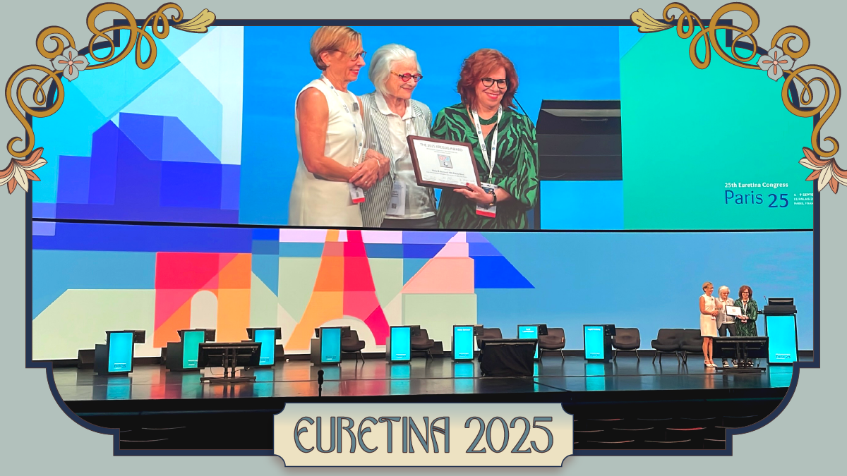
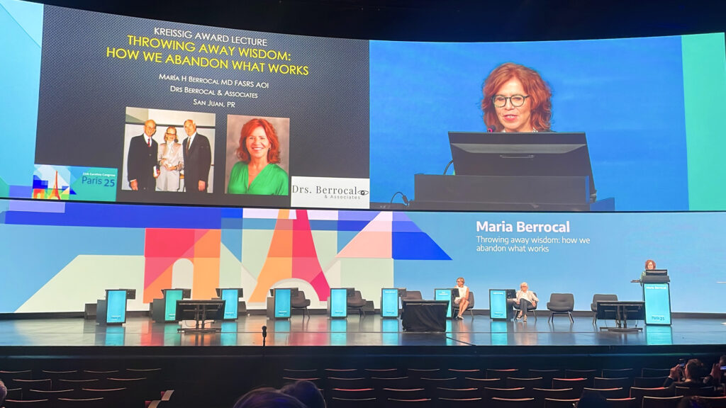
The Kreissig Award Lecture, named for Prof. Ingrid Kreissig (Germany), celebrates a lifetime of teaching minimalist, high-yield retinal surgery to generations of specialists. This year’s award went to Prof. Maria Berrocal (Puerto Rico), whose lecture was both a warning and a plea: Don’t abandon what has already stood the test of time.
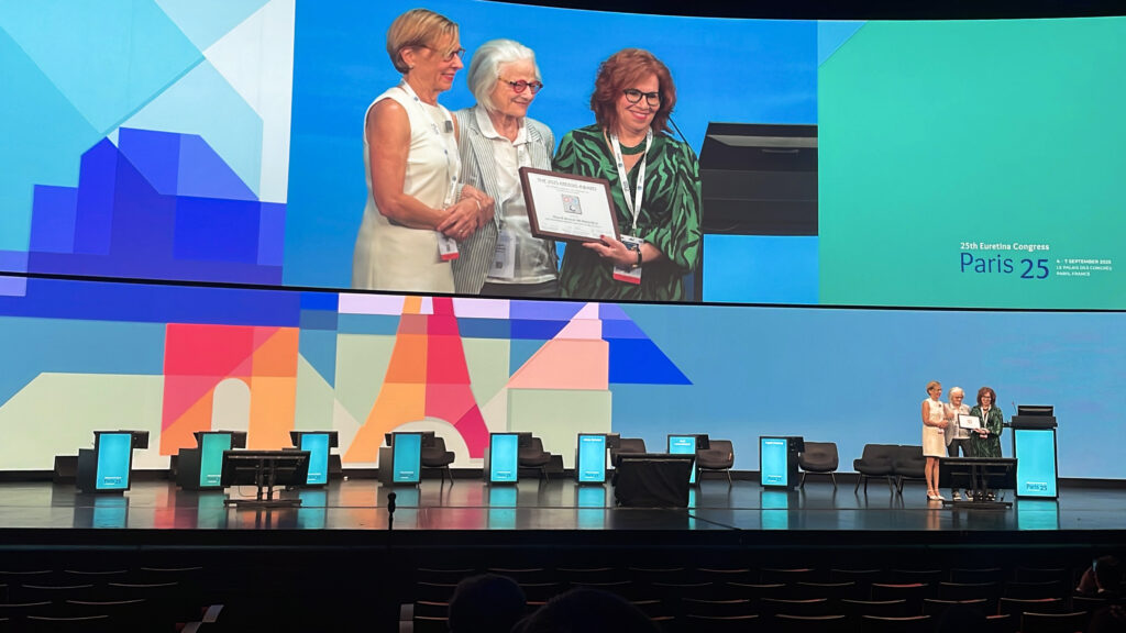
Presentation of the Kreissig Award to Prof. Maria Berrocal by Profs. Ingrid Kreissig and Anat Loewenstein
Her talk laid out the case for the field being too quick to sideline procedures that deliver lasting results with fewer complications.
Rediscovering the power of proven therapies
Prof. Berrocal began with rhegmatogenous retinal detachment, where she noted a single surgery success rate of 91 percent for scleral buckling versus only 84 percent for vitrectomy. The advantage, she emphasized, is even more pronounced in phakic eyes. Prof. Berrocal also pointed out that minimal segmental buckling without drainage consistently produces the strongest long-term outcomes with fewer complications than vitrectomy.
READ MORE: Latest Techniques for Retinal Detachment Success
To illustrate the point, she described a patient who had undergone both procedures—vitrectomy in one eye and a small buckle in the other. Both reached 20/20 acuity, but the postoperative journeys diverged sharply.
After vitrectomy, the patient endured nine weeks of disability; after the buckle, just two. The patient’s feedback was blunt: the second procedure was “fantastic,” because he could see immediately and did not have to endure head-down positioning.
Prof. Berrocal’s argument extended beyond detachment repair to an intriguing take on proliferative diabetic retinopathy (PDR): “Laser panretinal photocoagulation (PRP) is the standard of care for proliferative diabetic retinopathy.” PRP, she reminded the audience, induces durable regression that can last a decade or longer.
Anti-VEGF therapy, by contrast, only suppresses neovascularization temporarily and requires “continuous treatment forever.” That dependency, she warned, leaves patients highly vulnerable if they miss visits, and they often do.
READ MORE: A New Dawn for Retinal Care
The unsustainable cost of injections
The economic implications, too, are impossible to ignore. “In the United States, a quarter of all health money dollars go into treating diabetes complications. So if we just look at how many patients in the U.S. have proliferative diabetic retinopathy, we would require—just to treat proliferative diabetic retinopathy with injections—in one year, close to 4,000,000 injections. That’s over 300,000 injections monthly.”
And the challenge doesn’t stop at America’s borders. With diabetes rates surging worldwide, she warned that the burden of injections on health systems would be nothing short of catastrophic. As she put it plainly, in many regions such demand would “bankrupt any health system.”
For her, there was one overarching takeaway illustrated by her discussion of supposed vitreoretinal relics like buckling and PRP. Innovation, she argued, should be measured not in fashion or novelty, but in what endures for patients and health systems alike.
Gisbert Richard Lecture
The Gisbert Richard Lecture, named in honor of the legendary EURETINA co-founder and past president, recognizes outstanding contributions to vitreoretinal surgery and research.
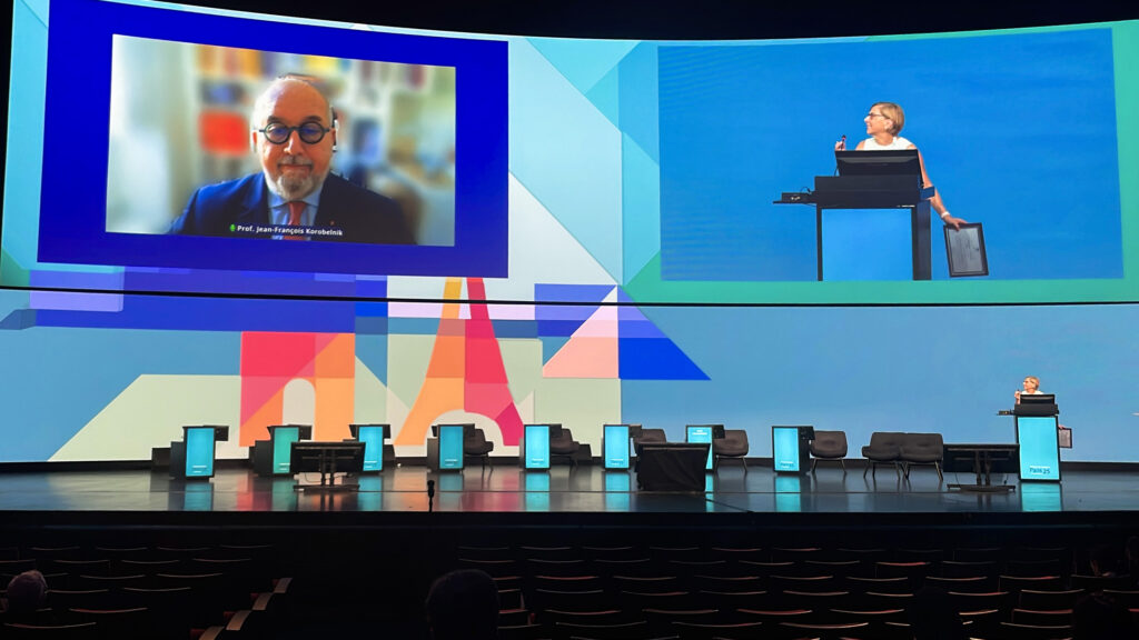
This year’s award went to Prof. Jean-François Korobelnik (France), a vaunted leader in retinal detachment and macular disease research. His lecture focused on the removal of intraocular foreign bodies.
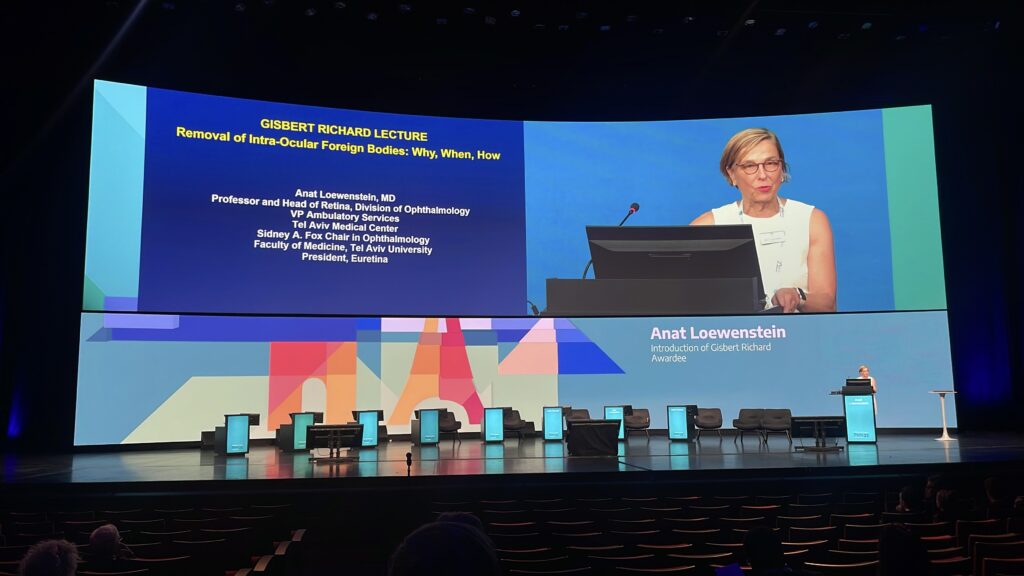
Prof. Anat Loewenstein on stage announcing the Gisbert Richard Lecture Award winner
Trauma tales and tips
He began succinctly with the first and most urgent step in trauma: “Just suture the wound. That’s all.” That initial closure, combined with intravitreal antibiotics (vancomycin and ceftazidime) forms the bedrock of prevention against endophthalmitis, according to Prof. Korobelnik. Systemic antibiotics, he noted, are no longer necessary. The goal is watertight closure to reduce infection and fibroblastic proliferation.
Imaging, too, follows a clear order: “The CT scan without contrast is definitely the modality of choice.” X-rays add little; ultrasound is invaluable when media are opaque or when wood or other non-metallic material is suspected. “MRI is contraindicated if a metallic intraocular foreign body is suspected.”
Timing, for him, is where judgment matters most. While immediate removal may be tempting, he prefers patience. “Most often, I recommend delayed removal, two, three to ten days after the initial surgery.” By then, hemorrhage settles, visualization improves and spontaneous posterior vitreous detachment may ease extraction.
READ MORE: Cracking the Code of Ophthalmic Trauma at APAO 2025
Mastering the “how” of extraction
Prof. Korobelnik continued with pearls on the removal of the foreign body. Under general anesthesia, he recommended performing a pars plana vitrectomy, induce PVD if possible, mobilize the foreign body gently, enlarge the sclerotomy and select the tool. While forceps, snares, and baskets still have roles, he underscored that “nowadays, we use the endocular magnet that will allow an easy lift of the foreign body at the surface of the retina, allowing its removal.”
His surgical videos revealed the patience required: shards that drift toward the disc, glass fragments that resist grasping, even a hidden piece of wood discovered only after careful probing. Each scenario demanded patience, adaptability and the ability to stay calm under pressure.
Yet even the most masterful surgery has limits. As Prof. Korobelnik reminded the audience, prognosis hinges on the initial damage: “If there is damage in the center of the posterior pole, the vision will be very poor.” Still, with orderly closure, targeted antibiotics, well-timed extraction and meticulous hands, many eyes can be saved—and many patients regain sight.
Surgical wisdom in two keys
Taken together, the two award lectures offered the audience more than surgical data; they offered perspective. Prof. Berrocal called for protecting proven, sustainable practices from being swept aside, while Prof. Korobelnik mapped out how discipline and precision can rescue vision when trauma threatens to take it away.
Both underscored that innovation in retina is not only about the newest tool or therapy. Sometimes, progress is remembering what works—and applying it with rigor.
Get the latest stories from the EURETINA 2025 here.
Editor’s Note: The 25th EURETINA Congress was held from 4-7 September, in Paris, France. Reporting for this story took place during the event. This content is intended exclusively for healthcare professionals. It is not intended for the general public. Products or therapies discussed may not be registered or approved in all jurisdictions, including Singapore.